Author: Irv Arons

A Most Memorable Trip: The Soviet Union Before the Breakup – The Interesting Adventures of A Consultant

Irv Arons

In the summer of 1990, my wife and I were invited to join a People to People Ambassador Tour to visit the Soviet Union. The USSR didn’t break up into its member states until the following year, late 1991. So, we got a look at what turned out to be a huge national facade – a strong military front and a “third world” infrastructure.

I was part of a medical group (medical laser specialists) that visited several hospitals and laser centers (?) within Moscow (Russia), Tbilisi (Soviet Georgia), Kharkov (Ukraine) and in St. Petersburg (again, in Russia). Fortunately, my wife was able to join up with a group of performing artists that were also on an Ambassador tour and, while we (the medical group) were doing our hospital visits, they visited their Soviet  peers,.

For the record, Moscow was bleak, with the exception of our visit to the Kremlin, and the gift shopping – my wife got a beautiful amber necklace from a street vendor for $20, and I came back with a set of matryoshka (nesting) dolls, with Gorbachev on top and Lenin deep inside at the bottom). In contrast, Tbilisi in Soviet Georgia was colorful and hot (90 degrees when we were there). The Ukraine, our next stop, was the food basket of Russia, but because they had few refrigerated trucks, there was no way of moving the food into the cities! Our final stop was St. Petersburg (or Leningrad, whichever you prefer), which was beautiful. It is a land of rivers and bridges – and the Hermitage, the most beautiful museum that we have ever visited!

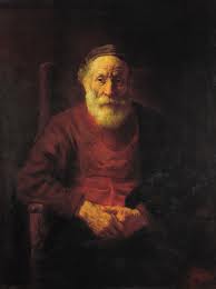
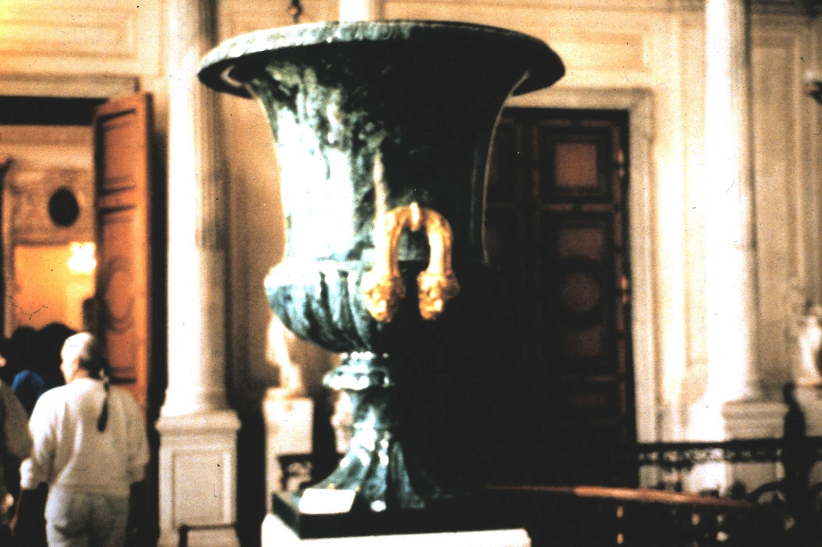
A brief anecdote – the day before our visit to the Hermitage, one of my colleagues ran out of 35 mm film and I gave him a couple of rolls of the 400 speed film that I was using. The next day, he replaced my rolls with a couple of rolls of 1000 speed film, which enabled me to take some beautiful pictures inside the Hermitage, including of the huge malachite urns and a couple of Rembrandt paintings (one of a man in a red coat – which I believe was a self-portrait, and Moses with his son Isaac on the mount) without needing to use a flash, which was forbidden inside the museum. I have included a few of these photos for your enjoyment – actually, the Rembrandt’s are from the web, as they are much better reproductions than I was able to achieve from my 35mm slides.)

We flew Aeroflot within the USSR – no seat belts, animals on board, and tea service served in cracked cups by heavy-set women with gold teeth! The planes were flown by military pilots, who were the first to get off, and the rear section of the plane was unloaded first to keep the planes balanced!

When we visited, most people worked for the state – and there wasn’t any work! So they would go to their jobs in the morning and, having nothing to do there, would be back on the streets by noon. It just goes to prove that the communist way of doing things didn’t work. There were no goods in the stores, and what we did see was both sparse and of poor quality (paper shoes?). And, long lines everywhere to get tobacco and vodka.

Based on my experiences, I published two articles about my trip; one about the state of healthcare (and lack of lasers) in the USSR, and a second on the state of eyecare in that part of the world. They are technical in nature but illustrate the differences between what the Soviet Union was telling the world and what we actually saw!

Here, for your further enlightenment, are the two articles I wrote about our (the medical groups’) experiences.

The State of Healthcare in the Soviet Union: The Lack of Medical Lasers
This article was published in both Medical Laser Industry Report, October 1990 and Laser Report, December 15, 1990.

Irving J. Arons
Arthur D. Little

During late July 1990, I was privileged to join a delegation of medical laser specialists and other health care professionals, under the auspices of the People to People Ambassador Program, invited to tour the medical community in the Soviet Union. Our group, organized by the American Society for Lasers in Medicine and Surgery, was composed of specialists in gynecology, plastic surgery, thoracic surgery, urology, general medicine, and myself, representing the field of ophthalmology. In addition to the ASLMS group, a veterinarian working with lasers, a health care safety specialist, and two medical technical/clinical lab specialists were included in our delegation.

We were able to visit a medical laser research institute in Moscow, and four hospitals, one each in Moscow (Soviet Russia), Tbilisi (Soviet Georgia), Kharkov (Soviet Ukraine), and Leningrad/St. Petersburg (Soviet Russia, again).

Our overall impression was that the facilities and equipment in use were woefully decrepit and/or non-existent or years behind Western standards. However, the medical personnel we met were dedicated professionals.

Of particular note, we found that the hospitals — even one claimed to be only three years old — were ill kept and falling apart. The facades were cracked and broken, the hallways and stairwells unswept, and the grounds surrounding the buildings not cared for at all. In counterpoint, we found the patient rooms were clean and staffed with dedicated nurses and doctors doing their best with what they had. This was particularly evident at the Karzigan Childrens Hospital in Moscow. The wards were filled with children with trauma of all kinds, but they were all smiling and very well cared for by an attentive staff of nurses and aides. It was here that we learned that a typical doctor with less than 10 years service earns between 240-260 rubles a month (the equivalent to about $40 at the official exchange rate of 6 rubles to the dollar, and only $20-30 at the black market exchange rate of 10-15 rubles to the dollar), a beginning research worker earns 140-150 rubles per month, and a surgical nurse get about 110 rubles for regular shifts. (In contrast, the bus drivers providing our transportation were paid 400 rubles/month and we were told that street sweepers earned as much as 700 rubles/month!)

The hospital equipment, especially for diagnosis and surgery, was particularly non-existent, and, according to our laboratory clinicians, the clinical laboratory equipment was barely adequate to care for the patients in the wards. But we must emphasize, both the hospital administration and medical staff were dedicated to providing the best care possible to their patients. What they lacked in equipment they more than made up with in numbers and dedication.

As for medical lasers, and new medical treatments using lasers, except for the laser institute in Moscow, the only lasers that the hospitals seemed to have were low powered therapeutic types, basically HeNes and GaAs infrared lasers. The hospital in Leningrad had several CO2 lasers, and claimed to have other surgical equipment, but we only saw therapeutic lasers in operation. We were told that only about 100 of approximately 15,000 hospital-based physicians in Kharkov have access to or use surgical lasers. We would guess that the percentages are not much different in the other 14 Soviet Republics. At the hospitals we visited, the majority of the half dozen lasers they claimed to have — if they had any — were either HeNe or GaAs therapy lasers, used to treat open sores, pain, and in one hospital in Tbilisi (Soviet Institute of Clinical & Therapeutical Research), for treating myocardial infarctions by clearing viruses in the blood through intravenous use of a HeNe laser connected to a fiber inserted through an arm vein. The same hospital also used a scanning HeNe laser to alleviate angina chest pain by scanning the laser beam across the patient’s chest!

At a trauma hospital in Leningrad (The Ambulatory Institute Hospital), we saw a 40-50 watt CO2 laser in the corner of an operating room, and another upstairs in a storeroom along with the usual HeNe lasers. We saw no evidence of a YAG laser although the laser specialist at this hospital claimed to have just received one, but which he had not yet unpacked. In the same storeroom with the spare CO2 laser, we were told that a small laser sitting on top of packing crates was a new UV laser, apparently solid-state since there was no evidence of any gas bottles or connections for one. (An interesting side note, we observed a burn therapy ward at this hospital, and it was disconcerting to see flypaper strips hanging from the ceiling.)

At the hospital in Kharkov (The Central Regional Hospital), we were told that they had an Ar laser used in ophthalmic treatments (but we did not see it).

The National Research Institute of Laser Surgery in Moscow claimed to be doing considerable research with medical lasers, performing 47,000 laser procedures annually. It is supposedly, one of 52 laser centers in the Soviet Union. We were given a presentation about the clinical research they were doing with five types of high power lasers, mostly CO2 and YAG — and two prototype free electron lasers, used for PDT studies. The FELs, according to the slides shown us, may be revolutionary, in that they appeared to be about the size of a large sized desk, much smaller than any other FEL I have seen — and I have seen the Stanford FEL and pictures of others. Attempts to find out more about this laser development were fruitless, however I plan to get back in touch with my contact at the Institute to see if I can possibly learn more about this exciting laser development. (Several weeks after my return to the States, I received a call telling me that a defector from the Moscow Laser Institute wanted me to know that he had built the laser in question and it was not an FEL, but rather an electron beam generator pumping a chemical laser use in their PDT work.) When we asked to see their lasers, we were politely told that they were in another building and couldn’t be shown to us. Apparently, the person holding the key to the lab was not available!

According to a profile of the Soviet healthcare community recently published by Medistat, a UK healthcare publication, what we saw in the Soviet Union this summer is typical and not out of line with what others have reported. The Medistat profile stated that the Soviet Union has some 23,000 hospitals with 3.6 million beds, and in addition, some 38,000 polyclinics and other outpatient centers. Capital investment in recent years has concentrated on the construction of new facilities to boost the number of beds, but many of the new facilities have been built at unsuitable sites, and the majority of Soviet hospitals having little in modern equipment with some lacking the basic necessities of adequate sanitary facilities or even heating.

The polyclinic is the main unit in the primary healthcare network, and the first point of contact for most Soviet patients. They serve districts of 50,000 to 60,000 inhabitants and are staffed by doctors responsible for around 2000 patients. In addition to general doctors, each polyclinic has specialists in area such as cardiovascular disease, oncology and renal medicine.

The Soviet Union has the highest number of doctors per capita, with a total of 1.3 million doctors serving a population of 275 million. In addition, there are 3.3 million medical assistants. Women doctors are well represented, accounting for about 70% of all doctors. (We were told the percentage was closer to 50%, at least at the facilities we visited.)

As you know, until recently, all planning was done centrally, and for five years in advance — the so called Five Year Plan. The most recent healthcare plan was put together in August 1987. That five-year plan attempted to address the chronic underfunding of the healthcare system, calling for new hospitals while neglecting older ones which will now need to undergo drastic major refurbishment. Expenditures for medical equipment had also been limited in the past, with the majority of funds allocated for new buildings. But the new policy included allocation of more resources for the purchase of modern equipment, in particular the polyclinic facilities were to be upgraded with proper diagnostic and treatment services to enable the patients to be treated at the clinic rather than to be referred to local hospitals. According to Medistat, 5.4 million rubles had been allocated for the purchase of new equipment over the two year period 1988-1989.

Discussing the state of the Soviet medical equipment market, Medistat states that the USSR suffers from a chronic shortage of equipment, particularly in the high tech areas such as computerized scanning, ultra-sound, renal equipment, and as we found out, laser treatment devices. Even basic equipment such as electrocardiographs and routine surgical instruments and disposable syringes are in short supply, with the problem being further aggravated by the fact that much of the Soviet produced equipment is sub-standard.

The current five-year plan envisages accelerating development in the medical industry and raising the technological level both within its industry and in its healthcare facilities. We hope that this can be accomplished. It is sorely needed.

In the report above, I discussed the state of healthcare in the USSR as we saw it in 1990. The hospitals and care appeared to be at least thirty years behind similar Western facilities, with a lack of instruments and supplies and crumbling buildings, but with dedicated medical personnel.

At the urging of a friend, I have added a brief piece, taken from Wikipedia, about how the healthcare system in the Soviet Union (and more specifically Russia) has changed since I was there – and from the way it is described below, it really hasn’t!

Healthcare in the Soviet Union and Russia
Source: Wikipedia

Pre-reform health care

Pre-1990s Soviet Russia had a totally socialist model of health care with a centralised, integrated, hierarchically organised with the government providing free health care to all citizens. All health personnel were state employees. Control of communicable diseases had priority over non-communicable ones. On the whole, the Soviet system tended to primary care, and placed much emphasis on specialist and hospital care.

The integrated model achieved considerable success in dealing with infectious diseases such as tuberculosis, typhoid fever and typhus. The effectiveness of the model declined with underinvestment. Despite the fact that the quality of care began to decline by the early 1980s,medical care and health outcomes were on par with western standards. Despite a doubling in the number of hospital beds and doctors per capita between 1950 and 1980, the lack of money that had been going into health was patently obvious. Some of the smaller hospitals had no radiology services, and a few had inadequate heating or water. A 1989 survey found that 20% of Russian hospitals did not have piped hot water and 3% did not even have piped cold water. 17% lacked adequate sanitation facilities. Every seventh hospital and polyclinic needed basic reconstruction. Five years after the reforms described below per capita spending on health care was still a meagre US$158 per year (about 8 times less than the average European social models in Spain, the UK and Finland, and 26 times that of the U.S. which spent US$4,187 at that time).

Reform in 1991-1993

The new Russia has changed to a mixed model of health care with private financing and provision running alongside state financing and provision. Article 41 of the 1993 constitution confirmed a citizen’s right to healthcare and medical assistance free of charge. This is achieved through compulsory medical insurance (OMS) rather than just tax funding. This and the introduction of new free market providers was intended to promote both efficiency and patient choice. A purchaser-provider split was also expected to help facilitate the restructuring of care, as resources would migrate to where there was greatest demand, reduce the excess capacity in the hospital sector and stimulate the development of primary care. Finally, it was intended that insurance contributions would supplement budget revenues and thus help to maintain adequate levels of healthcare funding.

The OECD reported that unfortunately, none of this has worked out as planned and the reforms have in many respects made the system worse. The population’s health has deteriorated on virtually every measure. Though this is by no means all due to the changes in health care structures, the reforms have proven to be woefully indequate at meeting the needs of the nation. Private health care delivery has not managed to make much inroads and public provision of health care still predominates.

The resulting system is overly complex and very inefficient. It has little in common with the model envisaged by the reformers. Although there are more than 300 private insurers and numerous public ones in the market, real competition for patients is rare leaving most patients with little or no effective choice of insurer, and in many places, no choice of health care provider either. The insurance companies have failed to develop as active, informed purchasers of health care services. Most are passive intermediaries, making money by simply channeling funds from regional OMS funds to healthcare providers.

National Projects

In 2006 a national project ‘Health’ was launched to improve the country’s healthcare system through improved funding and healthcare infrastructure. This plan helped equip hospitals and clinics with advanced, high-end equipment and ambulance systems, build new medical centers, as well as launch nation-wide vaccination programs and free health checks. The project has also been working on developing medical technology market through initiatives to blend healthcare and information technology. One of the focuses was made on salary increase of medical staff working in the primary care as well as their wider training programs.

The project was initiated by the Russian President Vladimir Putin and coordinated by the Presidential administration. It was mostly financed by the federal budget. However regional and municipal levels have also contributed a lot to the financing of the program.

Reform in 2011

After Vladimir Putin became president in 2000, there was significant growth in spending for public healthcare and in 2006 it exceed the pre-1991 level in real terms. Also life expectancy increased from 1991-93 levels, infant mortality rate dropped from 18.1 in 1995 to 8.4 in 2008. Russian Prime Minister Vladimir Putin announced a large-scale health-care reform in 2011 and pledged to allocate more than 300 billion rubles ($10 billion) in the next few years to improve health care in the country. He also said that obligatory medical insurance tax paid by companies for compulsory medical insurance will increase from current 3.1% to 5.1% starting from 2011.[27] Russia, anyhow, maintains several centers of excellence, such as the Fyodorov Eye Microsurgery Complex, founded in 1988 by Russian eye surgeon Svyatoslav Fyodorov. (See my next writeup,)

Eycare in the Soviet Union

This article was published in Vision Monday in August 1990.

Irving J. Arons
Arthur D. Little

I have just returned from a two week tour of four cities within the Soviet Union.  I had anticipated telling you about the state of eyecare within the USSR, but unfortunately, our visit to the premier Moscow Research Institute of Eye Microsurgery was canceled at the last minute, as we were informed by Dr. Fyodorov that the “Institute will be closed in July for preventative maintenance,” and that “your visit at the term (sic) you have stated is inexpedient.”  We had originally been invited following my February letter requesting to have our delegation visit the clinic. In fact, in late June we were told that we were welcome to visit the clinic, which was scheduled to shut down in August for holiday.

Apparently, the hotel we stayed at in Moscow, the Kosmos, is part of the Intourist program to provide visitors with eye care at Fyodorov’s Moscow clinic.  We found a brochure advertising “Beautiful Eyes for Everybody,” and describing how the Moscow Research Institute of Eye Microsurgery “treats 22,000 people annually, restoring or improving vision and removing the need to wear eyeglasses for many.”  In addition to RK (radial keratotomy), the clinic claims to do laser treatment of secondary cataract, laser treatment of glaucoma, and laser treatment of “complicated myopia of high degree.”  (It must be one of the only facilities with surgical lasers in the USSR, as we visited a laser research institute and three hospitals and saw very few surgical lasers — mostly therapeutic HeNe and GaAs biostimulation type devices.)

The brochure goes on to state that your pre-operative stay is arranged at the Kosmos, and following the outpatient surgery, your post-operative treatment is done at the hotel by a team of qualified doctors and nurses.  Foreign patients are offered a program of excursions, including theater tickets (the ballet only costs 4 rubles — the equivalent of 30 cents at the black market exchange rate) and other services offered by Intourist.  Fyodorov hopes to treat 20,000 foreigners a year by 1992.

A recent profile of Fyodorov in Fortune (May 1989) talked about the Fyodorov entrepreneurship, with his clinic being a $75 million a year business, and growing at 30% a year annually.  The clinics have over 5000 employers located at nine treatment centers across the Soviet Union, and include two factories producing eyeglasses and surgical instruments.  (In addition, Dr. Fyodorov recently spent some $12 million outfitting an 11,000 ton “floating eye hospital” called the Floks, which travels from port to port in the Persian Gulf offering RK and other eye surgeries.)

Having missed out on the opportunity to visit with Dr. Fyodorov, I would like to offer some personal observations about eye care in the Soviet Union.  As previously mentioned, our People-to-People delegation of medical laser specialists visited Moscow (Soviet Russia), Tblisi (Soviet Georgia), Kharkov (Soviet Ukraine), and Leningrad (back in Russia).  We saw very few optical shops (none that were open) and only a small number of Soviet citizens wearing eyeglasses!  One ophthalmologist at a central hospital in Kharkov told me that 50% of the people need corrective lenses — similar to the percentages in the rest of the world — but we saw very few people wearing lenses.  If 5% of the people in the streets had glasses, that’s a lot.  This says that Western technology and entrepreneurship could provide a needed service in the Soviet Union if a way could be found to open (and stock) optical retail shops in the major cities.  However, you must remember that the average citizen only earns about 200 rubles per month (the equivalent of about $350/month at the “official” business exchange rate or about $10-15/month at the black market exchange rate of 10-15 rubles to the dollar).  Therefore, the price of eyewear would have to be low for the average person to be able to afford it — unless the health care system can be convinced to reimburse or pay for the glasses.  (The government health care system pays Fyodorov’s clinic the equivalent of $300 for each RK procedure carried out.)

If a Moscow McDonalds can generate hours long lines for a $5 “Big Mac”, why not $20-40 eyeglasses at a Moscow Lenscrafters or a Leningrad Pearle Vision Center?

A First Trip to Japan: The Interesting Adventures of A Consultant

Irv Arons

In the summer of 1979, I had been hired to conduct a survey of all of the major contact lens companies operating around the world, for a US company interested in finding a partner for its contact lens company. I had completed my interviews of the companies in the US and Europe, and now had to interview the two companies producing lenses in Japan. Since this was my first trip to Japan, I decided to combine business with pleasure and took my wife along to spend a week on vacation, visiting Tokyo and Kyoto, before I was to meet up with a colleague from the Arthur D. Little (ADL) Tokyo office, who would act as my interpreter and take me to my meetings in Osaka and Nagoya.

The first leg of the trip was a three-day contact lens conference being held in San Francisco. There, I met up with a friend who ran a contact lens practice in Hawaii. Since he had clients who were on the management staff of Japan Airlines (who would be flying us from San Francisco to Tokyo), he was kind enough to arrange for my wife and I to be upgraded to First Class for our flight to Tokyo. (That was our first experience flying internationally in First Class and we liked it so much that I decided to upgrade our return tickets – at a cost of $600 each, as I recall, to fly First Class on our return flight from Tokyo to Boston. An excellent decision, as it was about a twelve-hour flight.)

We arrived in Tokyo and managed to find our way via bus from Narita airport to Tokyo Central Station, and to get a taxi to take us to our hotel, the Okura, in central Tokyo and near the ADL Tokyo office.

Our first venture out of the hotel was to the downtown shopping district of Tokyo. We had learned from our travel guide books that the best place to eat was at the restaurants located within the major department stores, up on the fifth floor. What the guide books neglected to tell us was that very few people spoke English and since we spoke only a few words of Japanese, we were left with only hand gestures!

At least at the restaurant, there were pictures of the several dishes available, and by pointing to the ones we wanted, we had a reasonable chance of getting something we could eat. But the next dilemma, the meals were served in a stack of bowls, and of course, with chop sticks. Was the top bowl broth or soup, or to wash your hands? And, if it is soup how do you eat it without spoons? Finally, another customer, sitting at a nearby table, sensing our discomfort, took pity on us and gestured to hold the bowl to your mouth and sip from it. So, that problem was solved.

The next day, I wanted to walk to the ADL Tokyo office that I was told was close by, so that I would know where it was for the following week when I had to check in. The street signs were a complete mystery and I had no idea how to get from the hotel to the office, even though I had instructions and a map!

Two Japanese men, passing by, sensed my dilemma and offered to help. After showing them the address of the office, they were kind enough to walk us right to the building, a very generous offer from strangers. The second offer of kindness to strangers from the Japanese people.

We had planned on spending a few days exploring Tokyo and then take a trip via the bullet train to Kyoto, the Japanese shrine city. However, our plans got changed because of a chance meeting at a coffee shop that afternoon.

An older gentleman introduced himself to us, while we were enjoying our coffee (or tea, I don’t recall). It turned out that he was a retired military officer, and also a former member of parliament. He offered to act as our tour guide and show us the real Tokyo and then take us to see Mount Fuji.

Since our plans were flexible, we agreed to his offer and arranged to meet him the following morning at the coffee shop for a guided tour of Tokyo. We hired a taxi for the day and our new guide took us to see and walk through the Parliament building and showed us several historic sites around the city. We ended that day with a visit to the Kabuki theater, being taken in through the people’s entrance and were able to watch the show from the first balcony. Quite an experience.

We enjoyed the tour so much, we agreed to let him be our tour guide the following day and take us to visit Mount Fuji, taking the bullet train (as long as we paid his fare). However, the weather didn’t cooperate and when we got to the mountain, it was completely fogged in. On the way back to Tokyo, we had another interesting experience. Three young women were on the opposite seats from us on the train and attempted to engage us in conversation. They claimed to be English teachers (or maybe students studying to be teachers) and wanted to practice their English with us. To be honest, we could barely understand them.

Since that was our last day of sightseeing, our gentleman guide asked us to meet him at a local Chinese restaurant that evening for a farewell dinner and to meet a few of his friends from the Kabuki theater. Little did we know that he had arranged for us to get the bill at the end of the evening and I got hit with a $250 check! (Recall that this was 1979, and the exchange rate for the yen was quite in my favor, about 350 yen to the dollar, but this was still an expensive meal!) I guess that was his way of getting paid back for acting as our tour guide for the couple of days we spent with him.

The next day, I left my wife at the hotel and made my way to the ADL Tokyo office to begin the business part of my trip, to visit the two contact lens companies in Nagoya and Osaka. (No wives allowed on business trips in Japan!)

Quite an interesting experience for my first trip to Japan!

My First European Trip: The Interesting Adventures of A Consultant

Irv Arons

Recall that I had toured Europe in the fall of 1979, visiting the various contact lens companies that were based there, as well as interviewing notable doctors involved in their use, on an assignment to assess the contact lens companies in both the U.S. and Europe for a Japanese client interested in acquiring technology for starting a contact lens business in Japan.

This was my first trip to Europe and I kept a diary of my daily travels – the first and only time I have ever done so. It’s a good thing I did, because there were several interesting incidents that occurred that you might enjoy.

I flew from Boston to JFK, to pick up my flight to London. I arrived in London at 10 PM, and quickly realized a had made a big mistake. I had a fairly large piece of luggage (I would be gone for over a week.), and hadn’t taken my wheels! (This was the fall of 1979, before wheeled luggage was in general use.) I made it onto the subway and got into London from Heathrow and finally found my hotel, the Bristol. Except they didn’t have my reservation! Luckily they had one room left and I took it and went to sleep without supper, as the kitchen wasn’t open at that time of night.

The next morning, I met my ADL colleague from our London office, who would be my traveling companion (and translator). It turned out, the London office had made my hotel reservations at another hotel, and forgot to notify me. (Again, these were the days before email.) We then went on to the several meetings that we had arranged in the London area. I went to the final one that evening by myself – no problem – they speak Bloody English!

That evening my colleague had arranged for us to meet at a nearby pub for a dinner show. I was quite surprised to find that he and his girlfriend had brought along another girl, his girlfriend’s roommate, whose boyfriend was out of town. After the dinner and show, I got dropped off at my hotel (alone) because we had a busy next day – four interviews and then flying off to Paris.

After our interview in Paris, the next day, we flew on to Strasbourg and our drive to Freiberg for a contact lens conference. We drove through very beautiful farm land and took a local inland route to the Rhine river and across into Germany at a local crossing. The border guards stopped us, took our passports, and searched our luggage thoroughly. They were puzzled by my voltage converter (European to 110v for my electric shaver), but after about ten minutes, finally allowed us to proceed into Germany and we went on to Freiberg and the CL conference.

I spent the weekend on my own, as my colleague took the train to Frankfort to spend some time with friends, after buying me a ticket for my Sunday train ride. Since I speak no German (although understand a little based on my Jewish background), I somehow managed to survive the weekend since I knew the word for chicken (hundchen!).

When I got to the train station on Sunday morning, I couldn’t figure out which track the Frankfort train was on, but luckily, some English gentlemen saw my dilemma and helped me out. It turned out that they were the world famous Amadeus Quartet and also going to Frankfurt. I thoroughly enjoyed my train ride talking to them.

Arriving at the Frankfurt station, I met my ADL colleague and we took the train to our next stop, Aschaffenburg. We arrived early in the evening, and after dinner, went to a movie and I saw Moonraker in German and didn’t understand a word of it!

Following our nearly all day meeting in Aschaffenburg, we took a taxi to the Frankfurt airport for our flight back to London. After leaving my luggage to walk to the gate, I thought I heard my name over the loudspeaker, but since the announcement was in German, I wasn’t sure. Sure enough, when we arrived in London, I quickly discovered that my bag was still in Frankfort.

I didn’t panic too much, but since we were going out of London the following day for two days of meetings, I decided all I needed was a clean shirt and some deodorant to get me through until my bag showed up in London. I quickly went out to try and find a shirt and the only store open had a dress shirt that set me back 27 pounds (or about $50). Finally, about 8 PM that night I got a call that my luggage had made its way to London and would be delivered to my hotel room. You can be sure I got up early the next morning, before our trip out of town and returned that $50 shirt! (I kept the deodorant.)

After a few meetings in London, we took the train to Birmingham, had a couple of meetings there and on to Loughborough.

After several meetings, it was on to Hitchins and then Hempel Hempstead. (Yes, I did get to see quite a bit of England on that trip.)

The next day was my last in England and after several more meetings, I made my way via the subway to the airport and finally got on my flight back to the States.

After what I saw of the beauty of Europe, I made a promise that someday I would return. (And, my wife and I did. A few years later we took a sixteen day tour of eight countries – “If it was Tuesday, it must be Belgium!” As it turned out, we did travel through Belgium on our way from Paris to Amsterdam on a Tuesday.)

We have since gone back to Europe several times, particularly to Switzerland and to Spain, and even took a river boat trip down the great rivers of Europe, from Amsterdam to Vienna, passing through sixty-six locks along the way.

The Soft Contact Lens Industry and My Role in its Growth: The Interesting Adventures of A Consultant

Irv Arons

The Bausch & Lomb Soflens, the original soft contact lens, was first approved for marketing in March 1972, My first contact lens assignment began in June of that year, an assessment of the “safety and efficacy” of the Soflens. Over the following fifteen years, I led over 150 assignments involving both soft and gas permeable contact lenses, becoming, in that period, the “guru” of the contact lens industry.

But one of the most interesting assignments, in the fall of 1979, involved my traveling all over the United States and Europe to investigate possible technical partners for a Japanese company interested in entering the contact lens business.

In September of that year, after completing my U.S. visits, I traveled to Europe, visiting London, Paris, Strasbourg, Frieburg, Frankfort, back to London and finally, home to Boston to prepare a final report. (For some anecdotes from my first trip through Europe, see the following writeup – My First European Trip.) In November I traveled to Tokyo to present the results of my study. An Arthur D. Little (ADL) colleague and I flew to Tokyo via New York City on a Friday, arriving on Saturday. The following day, Sunday, I returned a phone call to another client who had left an urgent message that he desperately needed to speak with me and that I should call him at home over the weekend.

At 8 AM Tokyo time, which was about 7 PM (the previous day) in the U.S., I reached Steve Martin of Ciba Geigy (the other client) and he asked me the strangest question. He told me that I was the No. 1 contact lens expert in the U.S., and he wanted to know who was No. 2? Apparently, his Swiss parent company had heard my name from so many people that they wanted another name to get another view of the industry. I told him that there wasn’t anyone else doing what I did and he agreed. He had himself made many calls around the industry and had only gotten my name. He said that he was going to let the Swiss people know this and would call me the following Thursday and probably give me another assignment.

As it was, he called me back the following Wednesday while I was still in Tokyo (leaving the next day to return to the states), and told me that he needed me to travel to Germany the following Sunday to evaluate the technology of a German contact lens company (Titmus Eurocon) in which he was interested. (Since I was planning to recommend the same company to our Japanese client, I politely asked him not to buy them until, at least, I could deliver our final report to the Japanese client!)

So, I arrived home that Thursday, wrote and sent my proposal to Steve (Ciba Geigy) and met him at the Newark airport that Sunday prior to my overnight trip to Germany. I was part of a four or five men team (the others being optometrists and ophthalmologists) who were being sent to Germany to evaluate the German company’s technology.

We flew overnight, arrived in country early the next morning and immediately went to work at the German company. After spending the day in discussions, the company officials offered to take us out for dinner and entertainment. With all of my recent travels, I was literally out of it and  politely declined and went to bed – I understand the rest of the group partied hard into the night – I have no idea how!

To make a long story short, after spending about three days in Germany, I came home, wrote my report and later heard that it was the influential piece of Steve’s presentation to management that gave him the go ahead to acquire the German company’s technology and to start CibaVision in 1980. He became its first president.

There are a couple of other stories about my role in advising companies that entered into the contact lens business. Here’s another that had an interesting twist.

Besides Ciba Geigy (CibaVision), I was also involved in the technical assessment of Frontier Contact Lens in Jacksonville, FL, for Johnson & Johnson, which they later acquired, and which became Vistakon.

In an earlier assignment for J&J, I did an assessment of Wesley Jessen in Chicago, but they passed on that company. However, about six months later, I was asked by Schering-Plough to visit in New Jersey about their potential acquisition of Wesley Jessen. I dutifully told them that yes, I had prepared a report on WJ, but I was under a confidentiality agreement not to discuss it. Schering said come on down anyway.

Lo, to my surprise, when I got to their offices, I was presented with a copy of my report and asked to discuss it! My guess is that the president of WJ (Orrin Stine) had given them a copy, given to him by the people at J&J. (I recently asked Orrin, but he doesn’t recall having a copy of my report.) Since I now felt that the confidentiality agreement was no longer valid, I did discuss my report and Schering did acquire WJ, which, as I recall, they later sold to Ciba.

So, I guess I did have quite an impact on the soft lens industry, both in the States and other parts of the world.

One last case and I’ll call it quits.

In August 1987, in a landmark case in the Tax Court in Wilmington, Delaware, ADL was hired by Baker & McKensy to assist in their representation of Bausch & Lomb in an international transfer pricing case against the IRS. It seems that B&L had transferred its technology to Ireland, where it was manufacturing most of its contact lenses then shipping them to the rest of Europe and to the U.S. for sale. This reduced substantially the amount of income B&L reported in the U.S. and allowed them to enjoy a much lower tax in Ireland. The questions before the Tax Court concerned the appropriate transfer price for the contact lenses and the royalty B&L Ireland owed to its U.S. parent company.

I was B&Ls contact lens industry expert and my ADL colleague, Dr. Irving Plotkin, their expert on foreign transfer pricing and royalties. (In fact he frequently worked both sides of the aisle, sometime being an IRS expert and other times working for the taxpayers!)

In any event, we prevailed and with a split decision by the tax court (mostly in B&L’s favor), saved B&L in the order of $15 million for the tax years in question, and perhaps tens of millions for succeeding years. This case set a precedent that became the law in international transfer pricing cases.

It was quite an experience being on the witness stand and being cross examined by the Government’s lawyers. In fact, I was so excited being in that position that I inadvertently answered one of the lawyer’s questions with a “Yes sir!”, when she was obviously a woman, who was very pregnant, and within weeks of delivering her baby!

If you are interested in reading more about my work in the contact lens industry, additional information can be found on my blog: The Nature and Evolution of the Soft Contact Lens Industry in the United States (the title of my expert report in the B&L vs IRS case).

Articles Published in Ophthalmic Journals in 2015

In addition to the several updates posted on this blog in 2015 (see previous posting), I have prepared four articles that have appeared in ophthalmic journals this year.

Here is a brief summary of the four articles, including links to the online versions:
Regenerating the Retina – February 2015
This article, published in the February 2015 issue of The Ophthalmologist, describes the use of stem cell-derived retinal progenitor cells (RPCs), that are being investigated for reviving/repairing/rejuvenating damaged photoreceptors to bring back sight to those who have lost it due to a retinal degenerative disease, including choroideremeia (CHM), retinitis pigmentosa (RP), Leber’s congenital amaurosis (LCA) and Stargardt’s disease (Stargardt Macular Dystrophy – SMD). It discusses the four companies that are conducting clinical research, as well as the work underway at several university and institutional laboratories.
To read the complete write up, please use the following link: https://theophthalmologist.com/issues/0215/regenerating-the-retina/
Update: Since the article appeared, two of the companies discussed, jCyte and ReNeuron have either begun a clinical trial (jCyte), or announced the start of one (ReNeuron).
With the likelihood of a gene therapy and/or a stem cell treatment for retinal diseases to be approved for marketing within the next two to three years, it is time for the ophthalmic community – the suppliers, practitioners, patients and payers –  to start thinking about how much these regenerative medicine treatments are likely to cost, and how patients and the healthcare system will pay for them..
In The Economics of Gene Therapy, appearing in the May 2015 issue of The Ophthalmologist, I propose a pricing model for Regenerative Medicine in Ophthalmology, based on the population of patients to be treated, and suggest that an annuity program model, based on performance and duration of efficacy, could be used to pay for it.
Let the dialogue begin.
To read all about it, please follow this link: http://tinyurl.com/GeneTherapyCosts

An update of the latest clinical information in the use of gene therapy in treating several retinal diseases, including Leber’s Congenital Amaurosis (LCA); the wet form of Age-Related Macular Degeneration (wet-AMD); Choroideremia (CHM); Stargardt’s Macular Dystrophy (SMD); Retinitis Pigmentosa (RP); and Ushers Syndrome (US). Included is the proposed model of pricing for some gene therapy treatments, based on the population of patients likely to be treated.
To read the full article, published in the October issue of Retinal Physician, please follow this link: http://tinyurl.com/RetPhys-GT-Update

Optogenetics is the introduction of protein-based, light-activated chemicals into still functional retinal cells in the vision chain, that upon activation, send electrical signals along the optic nerve to the brain, providing rudimentary vision, that was lost with the death or damage of the photoreceptors.
This report, The Optogenetics Option, describes the efforts of four companies and ten universities, using either gene therapy, or other means, to deliver light-activatable proteins or chemicals (photoswitches) to still functioning cells within the retina (ganglion and/or bipolar cells) or, in some cases, the use of light activatable implants, that will deliver light signals to the brain to provide some rudimentary vision when the photoreceptors, that normally provide that function, cannot.
To read the article, published in the November issue of The Ophthalmologist, please follow this link: http://tinyurl.com/OptogeneticsOption

Blog Articles Published in 2015

2015 was another busy year in writing about new developments in treating retinal diseases. During the year, I published seven blog entries (and another four published articles – to be indexed separately). Here are the highlights of the blog writeups:
Since I last wrote about Iluvien, Alimera Sciences and pSivida have announced additional marketing approvals for its use in treating chronic DME. The product is now approved for use in fourteen countries, including the U.S. In addition, pSivida is about to begin a Phase III study of its Medidur device for the treatment of uveitis, which should lead to a fast-track to approval.
But the real reason for this update was the recently published story about how an eye doctor reached out to Paul Ashton, CEO of pSivida, to package an old HIV anti-viral, ganciclovir, in his drug delivery system, to save the sight of a person undergoing chemotherapy for leukemia, who also needed a bone marrow transplant. The chemo and radiation treatment for the bone marrow transplant weakened his immune system, preventing control of his cytomegalovirus condition that began attacking his retina. Instead of painful weekly anti-viral injections, the doctor sought to use Paul’s drug delivery system to systemically treat the virus in the eye.
To read the whole story and the results, and for the latest information about Iluvien, please follow this link: http://tinyurl.com/IluvienUpdate9
With the presentation of the three-year safety results of the INTREPID study (at EURETINA), to evaluate the safety and efficacy of the Oraya Iray Therapy in conjunction with as-needed anti-VEGF injections for wet AMD, and the recent collaboration agreement with Carl Zeiss Meditec, Oraya Therapeutics is well on its way in implementing its growth strategy in commercializing Oraya Therapy in Europe and, some day, in the U.S.
The company now has nine centers providing therapeutic treatment in Europe; four centers in the UK, one in Switzerland, and four in Germany.
To read more about this combination treatment to reduce the number of anti-VEGF treatments needed to control wet AMD, and the collaboration agreement with Carl Zeiss Meditec, please see: http://tinyurl.com/OrayaUpdate3
Avalanche Biotechnologies in Menlo Park and the University of Washington in Seattle announced a licensing agreement to develop the first gene therapy treatment for treating color blindness. The deal brings together a gene therapy technique developed by Avalanche with the expertise of vision researchers at the University of Washington.
In addition, Avalanche will incorporate research licensed from UCal Berkeley to deliver the gene therapy treatment non-surgically via an  injection into the vitreous, rather than into the retina.
To read more, please follow this link: http://tinyurl.com/AvalancheBio2

As Crystal Gayle sang in her smash hit, `Don’t It Make My Brown Eyes Blue’, now (soon) you will be able to permanently change your brown eyes to blue. A California company has come up with a laser procedure that will safely accomplish this in about 30 seconds per eye. Human clinical testing is underway and the company, Strōma Medical, hopes to have the procedure on the market (outside of the U.S. first) in less than two years, once the clinical trials are completed.
To read the rest of the story, please follow this link: http://tinyurl.com/brown-to-blue
My article about the use of lasers to treat floaters, written five year ago, remains the most widely read piece on my blog. I frequently am asked if I have updated the list of doctors who now use lasers to treat floaters – in addition to the three I profiled in my U.S. writeup (Using Lasers to Treat Vitreous Floaters: Laser Vitreolysis) and the six doctors in the UK/Europe piece (Using Lasers to Treat Vitreous Floaters: Laser Vitreolysis in the UK and Europe).
I took notice when Ellex Medical announced a new YAG laser specifically for treating floaters in the fall of 2012, and about six months ago, I decided to request a list of doctors who have obtained the laser, and were now treating patients’ floaters. Better than a list, Ellex decided, to respond to my request, by building an app to locate doctors using their laser and put it on their website. That app is now active and is applicable to doctors worldwide using the new Ultra Q Reflex YAG laser to treat floaters. As of June 30th, there are 58 physicians listed worldwide using this laser.
To read more about the laser and use the app, see my update: http://tinyurl.com/vitreolysis-Update
Some insight into the Spark Therapeutics Phase III clinical trial results and longevity data. In this report, the company’s principal investigator, Dr. Stephen Russell presented highlights of the initial findings in this important Phase III clinical trial, that could lead to the first gene therapy approval in the United States.
Basically, it was found that the injection of SPK-RPR65 did lead to increased functional vision for the treated patients, compared to the control subjects – and the effect appears to last for over three years, based on the original patients treated in the Phase I study.
Is this the “forever fix”, or a step along the way? To read the full story, please follow this link:  http://tinyurl.com/GeneTherapyUpdate22
Three German ophthalmological groups provided guidance for German ophthalmologists to better identify those patients with wet AMD that might benefit from the use of the Iray Stereotactic Radiotherapy System as an adjuctive treatment to the use of anti-VEGFs in the treatment of neovascular macular degeneration, based on studies conducted by the company to date.
To read the full story, please follow this link: http://tinyurl.com/OrayaUpdate4

Oraya IRay Update 4: Oraya Therapeutics Receives Guidance for Use in Germany

When I last updated the progress of Oraya Therapeutics, in January of this year (Oraya IRay Update 3: Oraya Now Operating at Nine European Centers and Partnership with Carl Zeiss Meditec), I noted the collaboration agreement between Oraya and Carl Zeiss Meditec to provide funding to Oraya over a period of up to two years for the implementation of Oraya’s growth strategy, and I reported on the three-year INTREPID safety results, presented the previous September. Here now is the latest information on the progress of the company to bring this adjunctive treatment for the wet form of AMD to the professions.
The German Ophthalmological Society, the Retina Society and the Professional Association of German Ophthalmologists (DOG) have issued a joint opinion on the adjunctive use of radiotherapy with Oraya Therapeutics’ IRay Stereotactic Radiotherapy System for wet AMD. Their opinion makes it possible for ophthalmologists throughout Germany to identify patients that can benefit from Oraya Therapy in conjunction with anti-VEGF treatment.
Oraya Therapy is currently commercially available at eleven sites in Europe, with more than 550 patients treated to date with Oraya Therapy in three European countries – Germany, Switzerland and the United Kingdom.
According to the opinion, which takes the study data published to date into account, adjunctive stereotactic radiotherapy of neovascular AMD with the IRay system can be considered on an individual basis. Some of the parameters that ophthalmologists should consider include:
●    If symptoms of choroidal neovascularization (CNV) activity such as intra-retinal fluid of bleeding are present, corresponding to a recommendation of VEGF inhibitors;
●    If the ongoing anti-VEGF therapy has taken place over a period of at least six months, thus ruling out undertreatment; and,
●    If despite intensive injection therapy, no charge in the activity state of the CNV is achieved and there is no reasonable expectation of a decrease of the required high frequency of retreatment for the future.
“The joint opinion now provides guidance for ophthalmologists throughout Germany to identify those patients who can benefit from Oraya Therapy for wet AMD. There is a large patient population that does not respond well to anti-VEGF monotherapy, and our aim is to offer these patients in Germany an additional option to maintain their vision and also decrease the burden of frequent injections,” said Oraya Therapeutics CEO Jim Taylor. “We are well positioned with our IRay system at major university eye clinics in Germany, and will continue to expand our presence to make the therapy available to the patients who qualify and elect to pursue this alternative.”
The full opinion, titled “Stellungnahme von DOG, RG und BVA zur Strahlentherapie bei neovaskulärer altersabhängiger Makuladegeneration” is accessible within the German Ophthalmological Society website at:  http://www.dog.org/wp-content/uploads/2015/10/Stellungnahme-RG-DOG-BVA-zur-Strahlentherapie-bei-neovaskul%C3%A4rer-altersabh%C3%A4ngiger-Makuladegeneration.pdf.
The IRay Radiotherapy system is a CE marked medical device. In the U.S., the IRay system is an investigational device and is not yet available for sale.
For any of you reading this for the first time, please refer to my original full report describing the IRay system: Oraya IRay In-office Stereotactic X-ray Treatment for AMD: A First Report, written in January 2009.

Gene Therapy in Ophthalmology Update 22: First Spark Therapeutic Phase III Clinical Trial Results and Phase I Longevity Data

My article updating the latest information on the status of gene therapy clinical trials, What’s New in Gene Therapy for Ophthalmology? was published in the October issue of Retinal Physician. The final draft was submitted just prior to this important news from Spark Therapeutics providing information about their Phase III clinical trial results.
Principal Investigator Stephen R. Russell, MD, of the Stephen A. Wynn Institute for Vision Research at the University of Iowa, presented Phase III data highlighting the rate, breadth and magnitude of changes following administration of SPK-RPE65 to patients with Leber’s Congenital Amaurosis (LCA), at the Retina Society Meeting held in Paris on October 10th.
In addition, Dr. Russell presented data on the three-year durability of improvements in the same measures of functional vision and light sensitivity in a cohort of subjects from the earlier Phase I trial.
This presentation built on top-line results of a randomized controlled multi-center Phase III trial previously announced by Spark on October 5, which demonstrated a highly statistically significant improvement in the intervention group compared to the control group in the primary endpoint and two of three secondary endpoints.
Significant Improvements and Strong Parallels in Mobility and Light Sensitivity Testing
Data presented highlighted a mean improvement in the functional vision of intervention subjects (n=20) of 1.9 specified lux levels, compared with an improvement of 0.2 specified lux levels in control subjects (n=9) as measured by the change in bilateral mobility testing (MT) between baseline and one year in the modified intent-to-treat (mITT) population. The mITT population (n=29) includes all subjects that received SPK-RPR65, and only those who continued beyond the baseline study visit. Two subjects in the intent-to-treat (ITT) population (n=31) that were randomized but never received SPK-RPE65 are excluded from this efficacy analysis population. Thirteen of the 20 subjects receiving SPK-RPE65 were able to pass the MT at one lux at year one, demonstrating maximum improvement measurable on the MT score. None of the nine control subjects followed was able to pass MT at one lux at year one.
In a corresponding finding in the first secondary efficacy endpoint, full-field light sensitivity threshold testing (FST*) for white light, intervention subjects demonstrated a highly statistically significant mean improvement of -2.06 log10 (candela second/m2) compared with decline of 0.04 log10 (candela second/m2) among control subjects (all analyses in mITT population).
(*FST is full-field light sensitivity threshold. As RPE65-mediated retinal dystrophies affect primarily rods, night blindness and loss of peripheral vision are typically the presenting symptoms; central vision may be relatively spared initially. That’s why FST was the first hierarchically-arranged secondary as opposed, for example, to visual acuity. It is more reflective of the underlying pathophysiology of the disease.)
Dr. Russell presented additional top-line analyses from the pivotal Phase III trial showing the rapid and sustained impact of SPK-RPE65 throughout the entire one-year study period. Significant differences emerged in both MT and FST by the first study visit, at 30 days. These effects were reproduced consistently at each subsequent study visit (at days 90, 180 and one year).
Dr. Russell said, “It’s exciting to see a consistency of improvement between the functional vision and visual function. The parallel effect seen in the prompt response in both the primary and first secondary endpoints highlights a critically important finding from the trial: that functional improvements measured through the mobility test change score correspond closely with the physiologic impact seen through FST.”
Figure 1: Phase III Trial of SPK-RPE65: MT and FST Over Time (mITT)

In addition, Dr. Russell presented data on the durability of effect after three years as measured by MT and FST in a cohort of subjects that participated in the Phase 1 open-label study, and would likely have met the eligibility criteria for the Phase 3 trial. All of these subjects continue to experience a durable improvement over three years from the time of administration to the contralateral, or second eye, with observation ongoing. These subjects received the same dose and volume of SPK-RPE65 that was used in the pivotal Phase 3 trial. The figures below reflect data from all subjects available for follow-up at each time point reported. Spark and the clinical investigators continue to follow study participants to evaluate the durability of response, and will provide further updates in the future through a series of peer-reviewed presentations and publications.
“We are pleased to provide these additional informative data, the totality of which highlight the rapid, sustained and durable effect associated with SPK-RPE65 across multiple functional and physiological parameters, at time points ranging from 30 days to three years,” said Jeffrey D. Marrazzo, co-founder and chief executive officer of Spark. “We will continue to analyze the data from our groundbreaking pivotal Phase 3 trial in order to further elucidate the potential positive, meaningful impact that SPK-RPE65 can have on the lives of patients with RPE65-mediated blinding conditions.”
Figure 2: Phase 1 Trial of SPK-RPE65: Durability of MT and FST Over Time

Pivotal, Phase 3 Trial Overview
The pivotal Phase 3 trial of SPK-RPE65 is the first successful randomized, controlled Phase 3 trial ever completed in gene therapy for a genetic disease. The multicenter trial randomized 31 subjects with confirmed RPE65 (LCA) gene mutations. The ITT population included 21 subjects in the intervention group and 10 in the control group.
For the primary endpoint, subjects were evaluated at multiple time points over the course of one year for their performance in navigating a mobility course under a variety of specified light levels ranging from one lux (equivalent to a moonless summer night) to 400 lux (a brightly lit office) using the bilateral testing condition. Each attempt was recorded, and the videos were sent to independent, centralized, masked graders to assign a pass/fail score based on speed and accuracy with which the subjects navigated the course.
In addition to the primary endpoint, the statistical analysis plan included three secondary endpoints tested statistically in the following hierarchical order:
●    FST (white light), which reflects underlying physiological function by measuring light sensitivity of the entire visual field.
    
●    Change in MT score for the assigned first eye, which compares the MT performance between baseline and year one for the first eye injected for the intervention group and, for the control group during the control year, the first eye injected after they crossed over.
●    Visual acuity (VA) testing, which measures changes in central vision by assessing the ability of the subject to read a standard eye chart.
A summary of top-line efficacy results follows:
Primary outcome (ITT)    
MT change score, bilateral      p = 0.001
Secondary outcomes (ITT)    
FST, averaged over both eyes               p < 0.001
MT change score, first injected eye      p = 0.001
VA, averaged over both eyes                p = 0.17
So What Does All This Mean?
In a story about the trial results published by Bloomberg Business News, the reporters commented, “A gene therapy maker showed this week it could make blind children and adults see. But the big question left is how long the effect will last.”
“Much depends on the answer, including how much the company can charge for the drug — a price tag some say could be more than $1 million a patient.”
The company has shown the effect has lasted for as long as three years without degradation of vision in some patients, providing back functional vision to those nearly blind children treated in the initial clinical trial.
According to Dr. Russell, the principal investigator, “Investors will look at the data and are going to go, `Wow this is the best data we’ve seen, not just in the eye, but on gene therapy, period’ and they’re right,”
Yet he cautioned that more information is needed before the drug can be considered a cure. While it produces a missing enzyme needed to sense light, it can’t restore light-sensing cells that have already died off due to this progressive disease. In the initial study results, released on October 5th, one standard measure of vision, visual acuity, didn’t improve by a statistically significant amount. (But, functional vision did!)
Spark also hasn’t finalized and published data on individual patients, and it’s possible that some responded better than others. “Age in particular may be a factor in how much a patient can improve,” said Dr. Russell. The company said in its statement that no serious adverse events have been seen so far in the trial.
So, the question remains, is this the “forever fix”, as author Riki Lewis has written in her book on gene therapy, or something less? Time will tell. But this is certainly an important step forward for the treatment of an inherited retinal disease.
References:
1.  What’s New in Gene Therapy for Ophthalmology?, Irv Arons, Retinal Physician, October 2015.
Stephen R. Russell MD, Retina Society Meeting, Paris, Stephen R. Russell MD, October 10, 2015.

5. ‘Holy Grail’ Seen as Gene Drug for Blind Produces Lasting Effect, Caroline Chen & Robert Langreth, Bloomberg Business News, October 10, 2015.

Using Lasers to Treat Vitreous Floaters: An Update

Since my first article on the use of a YAG laser to treat floaters (Using Lasers to Treat Vitreous Floaters: Laser Vitreolysis) appeared in this space in August 2010, it has become the most popular/read write-up that I have posted. About 10% of all visitors to my blog come to read that article – and I’ve had over 215,000 unique visitors. One of the most frequent questions I get asked is, is there anyone close to where I live that does the procedure? Since I haven’t kept track of who besides the three doctors I featured in that first write-up are now doing the procedure, and since Ellex Medical (Ellex) now produces and markets a specialized laser (the Ultra Q Reflex) specifically to treat floaters, I decided it was time for this update.
As described by Ellex, upon release of their new laser in the Fall of 2012, here are its features:
“Featuring Ellex’s proprietary Reflex light delivery system, our ophthalmologist customers can  use the Ultra Q Reflex YAG laser to treat floaters in a medically-reimbursable procedure, known as YAG laser vitreolysis,” according to Ellex CEO Tom Spurling.
While there are several YAG lasers on the market, none until now were designed specifically for the treatment of floaters.
“The Ultra Q Reflex is optimized for performing YAG laser treatments both in the anterior segment and posterior segment – making it ideal for the all conventional YAG laser treatments such as capsulotomy and iridotomy, as well as YAG laser vitreolysis for the treatment of floaters,” added Mr. Spurling.
Ultra Q Reflex for Nd:YAG Laser Vitreolysis

Floaters (vitreous strands) are small bundles of collagen fibers located in the eye’s vitreous, which cast visual shadows that impede quality of vision. Often considered benign, they are a result of degenerative vitreous syndrome (DVS; the natural breakdown and clumping of collagen in the vitreous) and posterior vitreous detachment (PVD; the age-related separation of the vitreous from the retina).

To date, surgical removal of the vitreous (vitrectomy) has been the standard approach to the treatment of floaters. Highly invasive, the procedure carries a significant risk of complications, such as infection, retinal detachment, macular edema, anterior vitreous detachment and residual floaters.

The proprietary slit lamp illumination tower design of the Ultra Q Reflex converges the operator’s vision, the target illumination, and the treatment beam onto the same optical path, and focuses them onto the same optical plane as the treatment beam. This minimizes the potential for focusing errors and the risk of damage to the natural lens or the retina — making the Ultra Q Reflex ideal for the treatment of floaters. In contrast, conventional YAG lasers offer a limited view of the vitreous, which can make it difficult to visualize vitreous strands and opacities.
I contacted Ellex Laser management and requested a list of the U.S. doctors who have purchased this laser and who now use it to treat their patient’s vitreous strands/floaters. The company, in response to my request,  has set up an application on its website, called “Find a Physician”.
By entering your location, a map will appear which will locate doctors near you that have access to this specialized laser and use it to treat vitreous floaters by performing laser vitreolysis. The link to the app is: http://www.floater-vitreolysis.com/find-a-physician/
PS: The app works worldwide!
(As of the date I checked – June 30th – there were 58 physicians listed worldwide.)
Good luck to all.
References:

A Laser Process for Changing Brown Eyes Blue

`Don’t It Make My Brown Eyes Blue’ was a smash hit song(1) for Crystal Gale in 1977. But it has been a wish for many a young girl with brown eyes (and even some older ones and perhaps some men) ever since. Over the years it has been possible to turn your brown eyes blue using contact lenses, first lightly tinted ones, which really didn’t do much for dark brown eyes, and later opaque tinted lenses, which would cover brown pigmented eyes and really turn them blue (or green or even other colors), although they created an opaque light eye, which does not exist in nature and thus looks contrived. There are even a couple of surgical procedures that can transform the iris, or enhance/darken the edge, but the question of safety quickly arises with any surgical procedure.
So, a week ago, when my wife told me what she had just seen that there was a laser process to turn brown eyes blue, on The View, I replied, “No way”. You see, I was a consultant to the medical laser industry, involved in the use of lasers in both ophthalmology and cosmetic surgery, for over twenty years and had never heard of lasers used in this way. Just to be sure, I Googled it, and much to my surprise, yes, a company, Strōma Medical Corporation, in Laguna Beach California, is developing a permanent, non-surgical laser procedure that will turn brown eyes blue!
I found an article written for Ophthalmology Business(2) that explained how they were doing it. It also listed a couple of well-known ophthalmologists – Perry Binder and Marguerite McDonald, who I know on a personal basis – that are involved with the company, on their Medical Advisory Board, thus assuring me that this business was legitimate. I got in touch with my ophthalmologist friends and they put me in touch with the Chairman and Chief Scientific Officer of the company, Dr. Gregg Homer, to learn more.
What I have learned is that the company has developed a unique, low intensity, highly specialized laser and diagnostic aiming system (with computerized iris mapping and tracking), that targets the brown pigment on the front surface of the iris, removing some or all of that pigment, thereby revealing the natural blue eye lying below.  (The blue in an eye is actually the result of the scattering of white light entering the iris, by tiny grey collagen fibers called “stroma”, and the reflection of the shorter blue light, similar to the light scattering of sunlight by atmospheric molecules that makes the sky appear to be blue.)
A half treated eye (for illustration purposes).

As explained by Dr. McDonald in the Ophthalmology Business article, describing how the procedure works, “This is a Q-switched neodynmium YAG laser, which produces a very highly discriminatory photo-absorbed frequency. The laser fires a series of small, computer-guided pulses across the iris, to photo-disrupt the stromal melanocytes (the brown pigment). Because of the photo-absorption properties of this laser, the energy passes through the clear cornea and it very selectively hits the brown melanocytes, leaving the cornea and the posterior iris stroma totally undisturbed. The photo-disrupted melanocytes release cytokine protein molecules into the anterior chamber and the cytokine signal recruits macrophages…that engulf and digest the photo-disturbed melanocytes as cellular debris.”

To put it in simpler terms, the laser beam “breaks up” the brown pigment in the front part of the iris into much smaller particles, similar to the way lasers are used to remove tattoos, by a process known as “selective photothermolysis”(3). This phenomenon, invented by Drs. John Parrish and Rox Anderson of the Wellman Laboratories of Photomedicine at Massachusetts General Hospital, uses the principle of delivering pulsed laser energy to a selected chromaphore within the target tissue, without damaging surrounding tissue.
Illustration of the iris in cross section, showing the anterior border layer, which is the target of the Strōma Medical process.
The smaller particles are then digested by the macrophages formed and eliminated from the iris and from the anterior chamber via the normal liquid outflow channels in the eye.
The Story Behind the Story
According to Dr. Homer, he became interested in the concept of changing eye color in the late 1990s, discovering a paper in the literature on iris pigmentation by RC Eagle Jr.(4) “I finally found that paper, which wasn’t available digitally, and I thought, ‘Now that we’ve done so much work with lasers on dermal pigment, it should be fairly easy to remove that iris pigment safely, which should, in turn, reveal a blue eye.” He went on, “Around 2001, I personally funded a small study at Cedars-Sinai Eye Institute in Los Angeles. We took brown-eyed rabbits and proved the concept, showing that we could change an eye’s color. These rabbits don’t’ have blue eyes, but what we showed is we could (safely) remove the pigment.”
In 2009, Dr. Homer and his colleagues raised $2 million in a Series A round to form the company, build a prototype laser device, and open a clinical trial. They achieved their goals, successfully treating 17 patients in Mexico with solid efficacy and no adverse events.

The Procedure
The Strōma Medical  procedure is non-invasive. It involves no incisions or injections of any kind. In fact, other than the use of a small device to help keep the patient’s eyelid open during the procedure and the application of a mild topical medication, there is little or no contact with the patient’s eye.
The patient sits in front of the Strōma  laser, and his or her head is stabilized. The patient is instructed to direct the untreated eye toward a tiny animation, located about one foot from the patient’s eye, while the procedure is completed. The procedure is then repeated to treat the other eye.
The Strōma Medical Laser work station.
The treating physician will inform the patient when he or she may drive and return to work. In most cases, the patient should be able to do so shortly after the procedure. For the first week or so following the procedure, the irises will get darker. Thereafter, they will grow progressively lighter, revealing the underlying natural blue color. The full color change process should take two to four weeks following the procedure.
Where Does the Process Stand?
According to Dr. Homer, the Strōma Medical laser process, which takes about 30 seconds per eye,  is still being clinically tested before being released commercially, first outside of the U.S.
To date, the company has completed preclinical studies on 50 Dutch-belted rabbits, and clinical studies on 17 human subjects treated in Mexico. The company is preparing its’ first large-scale pilot clinical study in Costa Rica, involving about 20 patients. Following the successful completion of that pilot study, the company plans to treat about 100 additional patients in multiple countries and follow them for a predetermined length of time. 
The company plans to release the product when it and the governing regulatory bodies are satisfied with the safety and efficacy of the procedure. Due to the relative cost and complexity of releasing a cosmetic medical device in the United States, they expect to release the procedure outside the United States first. The order of release in the non-U.S. territories will depend upon the market demand and regulatory environment in each territory.
Remaining Questions
The technique appears to be non-invasive, safe, and  painless. The laser treats only the front of the iris and does not enter the pupil or treat any portion of the inside of the eye where the nerves affecting vision are located.
But several questions do remain:
1.What happens to the pigment debris that leaves the iris? Will that debris clog up the normal drainage channels in the front of the eye, which can in turn cause glaucoma?
Strōma Medical claims that the digested particles released by the process are too fine to cause glaucoma and can easily pass through the trabecular meshwork and out into the anterior chamber — and that even if there were any complications, they would be short-term and easily remedied, including the use of a standard laser procedure – selective laser trabeculoplasty (SLT), commonly used to eliminate pigment from the trabecular meshwork that might contribute to a rise in intraocular pressure. However, a small risk still remains, and the remaining studies are intended to address that risk prior to commercial release.
2. Don’t the pigmentary layers of the iris provide ultra-violet and infrared light protection to the lens and the retina in the back of the eye? Won’t removal of one of the layers increase the likelihood of cataracts and increased retinal problems due to increased ultra-violet and infrared light exposure?
Dr. Homer acknowledges that naturally light eyes tend to be more sensitive to bright light. He explains, however, that light eyes are not only less pigmented on the front surface of the iris, but throughout the eye, including the retinal nerves in the back of the eye that respond to light.  Light sensitivity, he maintains, is the result of less retinal pigment, not less pigment on the front surface of the iris.  Because the procedure is limited to the removal of pigment from the front surface of the iris, its removal would not increase light sensitivity. Instead, the procedure is able to achieve something that nature does not – a light eye without light sensitivity.  
3. How much will the procedure cost?
According to Dr. Homer, the procedure would likely cost about $5,000 for both eyes in the U.S., but the physicians doing the procedure would set the price, not the company itself, and it would likely vary depending upon territorial demand curves and currency fluctuations.
4. What will be the costs to the ophthalmologists?
The financial model for the procedure will follow LASIK – the laser device (which will be sold or leased), a maintenance contract for the device, and a per-procedure fee. The pricing has not yet been determined, but the company expects the device and service contract to cost less than those for LASIK, and the per-procedure fee to cost more.
5. Have you estimated the size of the market? Perhaps using the tinted contact lens market as an example?
According to market research done by the company, the relevant potential worldwide market for manufacturers of eye color changing products will be those people who have dark eyes (brown or hazel), are affluent enough to afford the procedure cost, and are 20-60 years of age. When the market is mature, 10.2 million people worldwide could have their eye color changed each year.  The largest patient population for the Strōma Medical’s system may be in India and China, followed by Central and South America, Southern Europe, Japan, Korea, the Middle East, and the United States.
Further, the company believes that the market for permanent eye color change will be the fastest growing segment of the aesthetics market over the next ten years. The market will be made up of individuals who have the money and the desire to change their eye color. The annual sustainable, and achievable market opportunity for companies in this space is estimated to approach $2.9 billion in five years. For physicians and their clinics, the market opportunity could be $9.3 billion. Early adapters of the technology will come from the 25 million patients who currently wear colored contact lenses, and the estimated 70 million patients who have stopped wearing them for aesthetic, comfort or an adverse response reason. But, the market could be much larger as the consistency of the results and a positive safety profile is confirmed through solid clinical studies.
6. Finally, do you have any IP protection? What’s to prevent a laser manufacturer to build both a laser and a diagnostic and aiming system to compete directly with you – in fact, isn’t there an entity in Barcelona that is now providing the service?
The company has an extensive international patent portfolio, which includes international patents covering several critical elements of the laser device and patents in the U.S., Singapore, and Australia covering any method or system using any form of electromagnetic radiation to alter iris color. The company also has additional protection covering various ancillary features of the procedure, such as its proprietary iris mapping and tracking technology, scan pattern, and beam profile.
The company claims that the physician in Barcelona was using an off-the-shelf laser designed for laser iridotomy and posterior capsulotomy, which is ill-suited for iris color change. As a result, many patients were injured – the local medical societies opened an investigation, and the physician appears to have discontinued the procedure.
To obtain more information about the process and the Strōma Medical laser, please visit the company’s website at this link.

References:

1. Story Behind the Song: ‘Don’t It Make My Brown Eyes Blue’
 
2. Brown to blue: Procedure changes eye color, Erin Boyle, Ophthalmology Business, July 2013

3. The theory of selective photothermolysis predicts that the selective thermal damage of a pigmented target structure will result when a sufficient fluence at a wavelength preferentially absorbed by the target is delivered during a time equal to or less than the thermal relaxation time of the target. (“Selective photothermolysis: Precise microsurgery by selective absorption of pulsed radiation”, RR Anderson and JA Parish, Science, 220:524-527, 1983.)

4. Iris pigmentation and pigmented lesions: an ultrastructual study, Eagle RC Jr., Tran Am Ophthalmol. Soc., 1988;86:581-687.

5. Strōma Medical Website.

Avalanche Update 2: Avalanche and Univ. Of Washington Collaborate to Defeat Color Blindness

Avalanche Biotechnologies and the University of Washington Enter Into Exclusive License Agreement to Develop Gene Therapy Approach to Treat Color Blindness
As reported by NPR’s health blog, Shots, on March 25th, Avalanche Biotechnologies in Menlo Park and the University of Washington in Seattle announced a licensing agreement to develop the first treatment for colorblindness. The deal brings together a gene therapy technique developed by Avalanche with the expertise of vision researchers at the University of Washington.
“Our goal is to be treating colorblindness in clinical trials in patients in the next one to two years,” said Dr. Thomas Chalberg, the founder and CEO of Avalanche. The company will be entering IND-enabling studies this year.
The agreement has its roots in a scientific breakthrough that occurred six years ago. That’s when two vision researchers at the University of Washington used gene therapy to cure a common form of colorblindness in squirrel monkeys.
“This opened the possibility of ultimately getting this to cure colorblindness in humans,” says Jay Neitz, who runs the Color Vision Lab at UW along with his wife, Maureen Neitz.
The company also announced, that outside of the scope of the license agreement, Drs. Jay and Maureen Neitz, faculty in the UW’s Department of Ophthalmology and color vision deficiency (CVD) experts, have joined its Scientific Advisory Board. They will be technical advisors to the company on the science of vision.
“This agreement with the University of Washington and world-renowned vision scientists Drs. Jay and Maureen Neitz will help us further advance our goal of developing therapeutic products for the millions of people who suffer from CVD,” said Dr. Chalberg. “Our proprietary technology enables us to target the retina through intravitreal adeno-associated virus delivery, presenting, for the first time, the opportunity to pursue previously untreatable ophthalmic conditions such as CVD.”
Avalanche will build on gene therapy research conducted by the Neitz research team, who used gene therapy to confer color vision in two adult male squirrel monkeys that had been color blind since birth. This groundbreaking work demonstrating proof-of-concept for treating CVD was published in the journal Nature (1).
Curing color blindness involves delivering new genes to cells in the retina that respond to color. That’s how Jay and Maureen Neitz cured the squirrel monkeys six years ago. But the technique they used involved a surgical procedure on the retina.
For people, they desired a nonsurgical approach, something that had eluded researchers for years. Then a team at the University of California, Berkeley found a way to deliver genes using a simple injection into the vitreous, the clear gel that fills most of the eyeball.
Avalanche Biotechnologies has been working to improve and commercialize the Berkeley technique, said Chalberg.
So, Avalanche will combine the CVD gene therapy approach discovered by Drs. Jay and Maureen Neitz, with the licensed technology from the University of California at Berkeley and improved by Avalanche which will allow treatment via an intravitreal injection, similar to how wet AMD treatments are currently delivered in ophthalmologist’s offices, in a simple in-office procedure.
(I described this latter new virus vector delivery system in an article written in June 2013 entitled: Gene Therapy in Ophthalmology Update 19: A New Virus Vector for Safer Delivery of Gene Therapies)
According to Dr. Samuel Barone, Avalanche’s Chief Medical Officer, in response to my query if they were going to use the specific Berkeley vector research that I wrote about in Update 19, Dr. Barone said, “We licensed that intellectual property from Berkeley, and it serves as the basis for the delivery.  However, we have improved and optimized the technology, in better cone-specific transduction and protein expression.”

About CVD/Color Blindness and Avalanche’s Targeted Development Program
Color vision deficiency (CVD), also known as red-green color blindness, is among the most common genetic diseases. CVD affects approximately 8 percent of males and 0.5 percent of females, or more than 10 million people in the U.S alone. CVD is a visual impairment that impacts many aspects of everyday life, resulting in limitations in professional choices, compromised health and safety, and the inability to perform many activities of daily living.
A simulation from the Neitz lab of what color blindness looks like, with normal color vision on the left and red-green color blindness on the right.
Photopigments in the retina are crucial for perceiving color. People with normal color perception have three different types of photopigments. These photopigments are tuned to perceive either long wavelengths (red), middle wavelengths (green) or short wavelengths (blue), respectively.

The most common forms of CVD are due to genetic defects that lead to missing either the L-opsin (Type 1, or protan defects) or the M-opsin (Type 2, or deutan defects). Affected individuals have trouble distinguishing between red and green and between colors that contain red or green hues.
Ishihara plate, a typical test for red-green colorblindness.
Avalanche has two drug candidates targeting these areas. AVA-322 carries the gene for L-opsin and is being developed for the treatment of Type 1 color vision deficiency. AVA-323 carries the gene for M-opsin and is being developed for the treatment of Type 2 color deficiency.
Avalanche expects to file an IND in the second half of 2016 to support advancement of these drug candidates into clinical trials, using Avalanche’s breakthrough non-surgical intravitreal injection method to deliver genes directly to cone cells at the back of the eye. The company recently launched www.colorvisionawareness.com for patients with color blindness to receive information about the condition and potential research study opportunities.
To read more about Avalanche Biotechnologies and its research programs, please see:
Reference
1. Gene therapy for red-green colour blindness in adult primates, Mancuso, K. et al, Nature, 2009; 461:784-787.

Oraya IRay Update 3: Oraya Now Operating at Nine European Centers and Partnership with Carl Zeiss Meditec

The last time I checked in on Oraya, in March 2013 (Oraya IRay Update 2: INTREPID Two-year Results Meet Primary Clinical Endpoint – Results in At Least 35% Fewer anti-VEGF Injections), the company had just announced the two-year results of the INTREPID clinical study, showing favorable results in requiring fewer anti-VEGF treatments to treat wet AMD.
With the recent announcement (January 7, 2015) of a collaboration agreement between Oraya and Carl Zeiss Meditec, to provide funding to Oraya over a period of up to two years for the implementation of Oraya’s growth strategy, and a report of the three-year INTREPID safety results last September, I decided to publish this update.
First, the three-year INTREPID trial safety results.
The three-year safety results were presented on September 13, 2014, at a EURETINA seminar where physicians also discussed their clinical experiences treating patients with Oraya Therapy.
The three year safety evaluation consisted of detailed image analysis looking for the presence of microvascular changes related to radiotherapy. Although small localized changes were identified by the reading center in a quarter of the patients, they did not significantly affect vision.
Commenting on the INTREPID study, Professor Ian Rennie, Consultant Ophthalmologist and Professor of Ophthalmology and Orthoptics and International Expert in Treatments of Ocular Cancers at Sheffield Teaching Hospitals NHS Foundation Trust said, “Of the cases I reviewed that were identified by the study expert panel as having microvascular changes attributable to radiation, most cases were so subtle that I would consider them clinically insignificant.”
The INTREPID study was the first to evaluate the safety and efficacy of Oraya Therapy in conjunction with as-needed anti-VEGF injections for patients with wet AMD, and is the only sham-controlled double-masked trial to assess stereotactic radiotherapy for wet AMD. The study met primary and secondary endpoints and showed that Oraya Therapy significantly reduces the need for anti-VEGF injections while maintaining vision in the presence of a favorable safety profile. A total of 21 sites in five European countries participated in the trial. Two-year results showed that a broadly inclusive cohort of previously treated Wet AMD patients continued to receive the benefits of a 25 % mean reduction in anti-VEGF injections over two years. Additionally, the targeted patient population maintained an impressive 45 % mean reduction in injections through the two-year visit, with stable vision.
In simpler terms, as explained by Oraya President and CEO Jim Taylor, “The INTREPID trial enrolled a broad spectrum of patients, with some diagnosed out to 3 years prior (average ~15-18 months), and including many who’s disease process had progressed to central scarring and other related conditions. In this broad cohort, we saw the 25% reduction of injections with equivalent vision outcomes to the controls.”
Further, “In a subset of patients who had active leakage and lesions that had not grown so large as to extend outside the Oraya treatment zone, we saw the far more significant 45% reduction; and some suggestion of potential for positive vision benefit as well. This becomes our “target patient population” for commercial/clinical use, and it is important to note that our research suggests these patients make up 60%-70% of the patient population under wet AMD management in a typical clinic.”
These impressive results coupled with increasing interest from physicians and patients, have led the Heart of England NHS Foundation Trust to become the second NHS center to install the technology. Patient treatment at the Trust was due to commence in October 2014. The Heart of England closely follows the Royal Hallamshire Trust in Sheffield, which began offering this innovative therapy in July 2014. The commercial use of Oraya Therapy has been rapidly expanding in Europe, with the treatment available in a total of eight centers (see note below) across the United Kingdom, Germany and Switzerland. It is covered by insurance in all three countries.
“As we conclude this ground-breaking three-year study of Oraya Therapy, results are showing that treatment can, and is, maintaining patients’ vision while significantly enhancing their quality of life,” said JimTaylor. “This result, coupled with the rapid expansion of the availability of the Oraya Therapy in the UK, and indeed across Europe, makes this a very exciting time for the company.”
(Editor’s Note: As of January 2015, there are now nine centers treating patients; four in the UK, one in Switzerland, and four in Germany. See the company website for additional information.)
On January 7, 2015, Carl Zeiss Meditec AG and Oraya Therapeutics, Inc. (Oraya) jointly announced that the companies had entered into a collaboration agreement under which Carl Zeiss Meditec will provide funding to Oraya over a period of up to two years for the implementation of  Oraya’s growth strategy, and in turn receive rights in the company reaching up to a majority stake after two years.
The Oraya Therapy is available commercially in Germany, the UK and Switzerland, and the collaboration is intended to accelerate and expand these initial European market developments. While specific terms of the agreement were not disclosed, the companies noted that Carl Zeiss Meditec will be making a meaningful strategic investment in Oraya, and that further opportunities to leverage the companies’ respective technical and market expertise and resources will be reviewed.
In discussing the transaction, Dr. Ludwin Monz, President and CEO of Carl Zeiss Meditec AG, stated, “The current pharmaceutical treatment regimens for wet AMD are exceptionally costly and burdensome, and Oraya’s unique therapy offers a significant potential to positively impact the management of this debilitating disease.” He went on to add that, “ZEISS has a long tradition of bringing new and innovative technologies to the ophthalmic market, from the earliest slit lamps, to category leading products such as glaucoma field analyzers, Optical Coherence Tomography (OCT) and innovative femtosecond laser platforms. These types of technology innovations, all offering significant provider and patient benefits, are part of the core strategy of Carl Zeiss Meditec AG.”
Commenting on the agreement, Jim Taylor, CEO of Oraya, stated, “It is exceptionally rewarding to have the support and validation that are inherent in this commitment by Zeiss, a company universally recognized for its commitment to excellence in science as well as in patient-focused and physician-focused products and innovations. As a result of this collaboration, we have effectively enhanced our potential to positively impact patient outcomes, while significantly reducing the therapy burden for clinicians and providers as well.”
In the USA the Oraya IRay is still an investigational device and is not yet available for sale.

Iluvien Update 9: Additional Marketing Approvals; A New Ophthalmic Application; and An Interesting Human Interest Story

Since I last wrote about Iluvien back in September (Iluvien Update 8: Alimera Sciences Receives FDA Approval of Iluvien for Treatment of DME), when Alimera Sciences and pSivida announced the FDA approval of Iluvien for treating chronic DME, adding the U.S. to the approvals or pending approvals obtained in seventeen European countries – approved in thirteen and pending approval in an additional four others, the product has become commercially available in the UK and Germany and is scheduled to become available in Portugal shortly.
With the FDA approval, Iluvien should also become commercially available in the U.S. in early 2015.
In a recent news release, Dr. Paul Ashton, President and CEO of pSivida, said, “We continue to be pleased as Iluvien gains additional marketing approvals in Europe. We believe Iluvien’s efficacy and three-year duration make it an attractive treatment option for many DME patients, particularly in the U.S. where the drug has broader labeling.”

In addition to the application of Iluvien for the treatment of DME licensed to Alimera, pSivida is independently developing the product for the treatment of posterior uveitis. The FDA recently cleared the company’s Investigational New Drug (IND) application to treat posterior uveitis with its injectable, sustained-release micro-insert, permitting pSivida to move directly to a single Phase III trial under which it would enroll a total of 300 patients. The FDA is allowing pSivida to reference much of the data, including the clinical safety data, from the clinical trials of Iluvien for DME previously conducted by Alimera. Under the terms of its collaboration agreement with Alimera, pSivida has joint ownership of, and reference rights to, all clinical data and regulatory filings generated by Alimera, including its New Drug Application (NDA) for DME.
pSivida’s injectable micro-insert (Medidur) to treat posterior uveitis is a tiny tube about the size of an eyelash. It releases the off-patent steroid fluocinolone acetonide at a consistent rate over a period of approximately 36 months. The micro-insert is injected into the back of the eye during an office visit through the use of a fine gauge needle.
And, An Interesting Human Interest Story
What caught my attention, and brought me back to Iluvien (and the company, pSivida, that produces it and licenses it to Alimera Sciences for marketing) was a story that recently appeared in the Kansas City Star:
This is a story about a software engineer from Olathe, Kansas.
David Jiang was in danger of losing vision in his left eye when his eye specialist tracked down the inventor of a tiny eye implant that releases an anti-viral drug.

Jiang was examined recently by Siddhartha Ganguly (left) at the University of Kansas Cancer Center. 
David Jiang (left) says his vision remains slightly blurry but his retina is recovering after receiving the implant.
A year ago, after a routine physical, Jiang got the disturbing news that he had leukemia.
“I was shocked,” he said, when his doctor called him with the diagnosis. His life was in danger, the doctor warned him. Jiang had been heading out the door to lunch. He changed his destination to the University of Kansas Cancer Center. The next morning, he started chemotherapy.
But the chemo wasn’t doing the job of ridding Jiang’s body of his cancerous bone marrow and the abnormal white blood cells they were producing. Doctors decided he needed a bone marrow transplant, a tough and sometimes risky course of treatment. A bone marrow donor was found, and Jiang received the transplant in April.
So, what does this have to do with Jiang’s eyesight?
Before cancer patients undergo a bone marrow transplant, they must go through a rigorous course of chemotherapy or radiation to eliminate their own diseased marrow from their bones. The donated marrow is infused into their bloodstream and migrates to their bones, where it grows and begins producing healthy blood cells.
But bone marrow cells are also the source of the body’s immune system. Until new marrow can renew patients’ immune systems, they’re exceedingly vulnerable to all kinds of infections. And as with other kinds of transplants, there’s the potential for rejection, which can slow recovery.
When kidneys, livers and other organs are transplanted, the immune system of their new owner may sense them as foreign objects and launch a search and destroy mission. But with bone marrow transplants, rejection comes with a twist. It’s the bone marrow itself, the person’s new immune system, that sees its new home as the alien and attacks the patient. It’s called graft versus host disease.
That’s what happened to Jiang. “After the transplant, I was exhausted,” he said. “I had one complication after another.” There were headaches and stomach pain. To keep his graft versus host reaction in check, Jiang had to take drugs to suppress his immune system.
And that is how his eyesight was put in jeopardy.
Like most adults, Jiang is a carrier of cytomegalovirus, also called CMV. It’s scary-sounding, and the virus can be life-threatening. But people with healthy immune systems generally keep it in check and never know they carry it. But in people with compromised immune systems, such as those with AIDS or bone marrow transplants, cytomegalovirus can cause big trouble.
Depending on which organs CMV attacks, it can cause inflammation of the brain, seizures, coma, ulcers in the digestive tract, pneumonia or inflammation of the liver. The virus also can attack the eyes.
For Jiang, it began a painful attack on the retina of his left eye, blurring his vision.
“We needed to stop this virus where it was or it was going to eat away the center of his vision,” said Ajay Singh, Jiang’s eye specialist at the University of Kansas Hospital. Singh started twice weekly injections of an anti-viral drug (ganciclovir) directly into Jiang’s eyeball. The injections were painful and risked causing an infection. Singh couldn’t give Jiang an oral version of the drug because it could damage his new bone marrow.
But Singh knew that during the AIDS epidemic, CMV eye infections were so common that a tiny device was created that could be loaded with medication and implanted directly into a patient’s eye where it would slowly release the drug. Singh wanted one of these implants for Jiang – his patient was an ideal candidate – but they were nowhere to be found. It turned out that AIDS treatment had advanced so far that people with AIDS no longer were developing CMV eye infections. The implant manufacturer had pulled it off the market about a year or two ago, he found.
Singh was worried: The infection was a millimeter from the center of vision of Jiang’s retina. If he didn’t get an implant within a month, his vision could be lost. “The odds were stacked against him.”
Early in October, Singh started making calls to Singapore, India and Mexico City, searching for an implant. He emailed the CEO of the company that discontinued the device but never received an answer. Eventually, the people Singh was contacting told him of an eye doctor in Kentucky who knew the original inventor of the implant. The Kentucky doctor put the two in touch. And it is here that the inventor-entrepreneur enters the picture.
“Dr. Singh somehow rooted me out,” said Paul Ashton, CEO of Massachusetts-based pSivida. While pSivida had licensed its CMV implants to another company, it was still making other drug-dispensing eye implants.
Its newest is the size of an eyelash and can hold enough medication to treat diabetic eye disease for three years. Ashton offered Singh a slightly larger implant, about the size of a grain of rice, that would hold enough of the ganciclovir anti-viral drug.
“It was simply a matter of putting in a different drug,” Ashton said. “We were changing the ammunition but keeping the same gun.”
Singh recalled Ashton saying: “If you can send me the drug and get approval from the Food and Drug Administration, I will make you the implant.” So Ashton went to his laboratory, testing the permeable ends of the tiny tube to be sure they would allow the drug to ooze out at the right speed. “It was a month of many sleepless nights. It was a race against time,” Ashton said. “You can’t keep injecting someone in the eye. Something bad is going to happen.” Singh marveled at the speed with which Ashton’s laboratory worked. “It’s not like a car engine part” off the shelf, Singh said. “They have to test it. To do it in four weeks is a big deal.”
In early November, the implant was ready. Singh called the FDA for emergency approval for the implant. “The FDA responded at lightning speed,” he said. “I got an answer in four hours.”
On Nov. 5, Singh implanted the device in Jiang’s eye at KU Hospital.
“Now his cancer doctors can keep working on getting his bone marrow up and running,” Singh said. The implant will continue delivering the drug to Jiang’s eye for nine months to a year. “By that time, his immune system can recover and get the virus under control again.”
Jiang said his vision is still a bit blurry, but his retina is recovering. Singh has assured him that in a few months, with new prescription glasses, he will be seeing well again. So far, his cancer hasn’t returned and his graft versus host disease is well-controlled.
Jiang is grateful to the far-flung team that saved the vision in his left eye. “They all worked together. They worked so well together. I’m so fortunate.”
To read more about how the pSivida drug delivery system works, please take a look at my initial writeup of Iluvien: Iluvien and the Future of Ophthalmic Drug Delivery Systems.
Reference:

Tiny device and lots of teamwork save Olathe leukemia patient’s sight, Alan Bailey, The Kansas City Star, January 2, 2015.

Stem Cells in Ophthalmology Update 28: First Ophthalmic Stem Cell Treatment Recommended for Approval

On December 19th, the European Medicines Agency’s (EMA’s) Committee for Medicinal Products for Human Use (CHMP) recommended the stem cell product Holoclar (Chiesi Farmaceutici S.p.A.)  as a first-ever medicinal treatment for severe limbal stem cell deficiency, a condition caused by physical or chemical burns to the eye or eyes in adults, which can result in blindness.
The EMA has designated Holoclar as both an orphan medicine and an advanced therapy medicinal product, which enabled the manufacturer to receive free scientific advice and protocol assistance for drug development. The CHMP recommendation was based on an assessment by the expert Committee for Advanced Therapies. Such steps are taken to promote the development of medicines for rare diseases and to encourage innovative medicinal products, according to the EMA statement.
Holoclar is produced by Chiesi under an agreement with Holostem Terapie Avanzate, an Italian biotechnological company devoted to the development, manufacture, registration and distribution of Advanced Therapy products based on cultures of epithelial stem cells both for cell therapy and gene therapy. Holoclar was designed specifically for the treatment of severe limbal stem cell deficiency (LSCD).
The transparency of the cornea is essential to ensure the ability to see properly. Corneal cell renewal and repair are dependent upon the cells present in the limbus, which is found in a small area of the eye between the cornea and the conjunctiva.
Thermal or chemical burns to the eye can destroy the corneal surface (epithelium) and the limbus, causing a deficiency of limbal cells. If this happens, the cornea is covered by a different epithelium following an invasion of cells from the conjunctiva. This process leads to neovascularization, chronic inflammation and stromal scarring, rendering the cornea opaque and results in subsequent loss of vision. Conventional corneal transplants are an ineffective treatment in such cases.
Fig 1 Damaged eye before treatment
The therapy is based on cultures of limbal cells taken from the patient, which, once they have successfully grafted, regenerate the corneal epithelium and restore its functions. Limbal cell cultures even allow the possibility of treating patients with a loss of corneal epithelium in both eyes, provided that a small portion of limbus remains in one of the eyes.
Holoclar, is an autologous culture of limbal stem cells. It is made from a biopsy taken from a small undamaged area (minimum of 1-2 mm2) of the patient’s cornea and grown in the laboratory using cell culture, and transplanted in the affected eye or eyes after removal of the damaged area. Such cultures engraft and permanently regenerate a functional corneal epithelium allowing recovery of visual acuity. replacing damaged limbal stem cells.
Fig 2 A cultured sheet of corneal epithelium
Holoclar can offer an alternative to corneal transplantation for replacing altered corneal epithelium in some cases, and it has been shown to increase the chances of a successful corneal transplant where the injury has caused extensive eye damage. It reduces the risk of rejection compared with transplanting tissue from a donor and does not require surgery on the patient’s other eye as only a small biopsy is performed to collect the cells, thus reducing the risk of damage to the healthy eye. Therefore, Holoclar may also be suitable where both eyes are affected by moderate to severe LSCD.
Limbal stem cell deficiency is estimated to affect about 3.3 per 100,000 people in the EU, causing pain, photophobia, inflammation, corneal neovascularization, loss of corneal transparency, and eventually blindness.
The recommendation was made by the Committee for Medicinal Products for Human Use (CHMP) based on a robust assessment carried out by the Committee for Advanced Therapies (CAT), the Agency’s expert committee for ATMPs.
“This recommendation represents a major step forward in delivering new and innovative medicines to patients,” says Enrica Alteri, Head of EMA’s Human Medicines Evaluation Division. “EMA has used all available support tools to facilitate the development and assessment of Holoclar. It is an advanced therapy medicinal product that has been designated as an orphan medicine. This allowed the Agency to provide support including several rounds of free scientific advice to the applicant during Holoclar’s development.”
The Committee for Advanced Therapies and the CHMP panels determined that although the product’s benefits outweigh its risks, the marketing authorization should be conditional because the data thus far are retrospective and not yet comprehensive. Therefore, the EMA says, “an additional study on the use of Holoclar should be conducted.”
The opinion adopted by the CHMP at its December 2014 meeting is an intermediary step on Holoclar’s path to patient access. The CHMP opinion will now be sent to the European Commission for the adoption of a decision on an EU-wide marketing authorization. After that, decisions about price and reimbursement will take place at the level of each member state,  taking into account the potential role/use of this medicine in the context of the national health system of that country.
References:
1. First stem-cell therapy recommended for approval in EU, European Medicines Agency Press Release, December 19, 2014
2. First-Ever Stem Cell Therapy Recommended in EU, Miriam E. Tucker, Medscape, December 19, 2014

Stem Cells in Ophthalmology Update 27: ACT Interim Clinical Results Are Outstanding

Having treated 36 patients in two clinical trials for Stargardt’s Macular Dystrophy (SMD – 24 patients to date) and for dry Age-Related Macular Degeneration (AMD – 12 patients to date), Advanced Cell Technology reported the interim results obtained with 18 of these patients (9 in each trial) in the US-based studies. Both trials (NCT01345006 – Stargardt’s, and NCT01344993 – AMD) began in July 2011, giving the company up to three-year’s data for the earliest patients, and a median of 22 months followup for all. The interim results were reported in The Lancet, published online October 14, 2014 in: “Human embryonic stem cell-derived retinal pigment epithelium in patients with age-related macular degeneration and Stargardt’s macular dystrophy: follow-up of two open-label phase 1/2 studies”.
An additional Stargardt’s trial, being conducted at two clinics in the United Kingdom, with 12 patients enrolled (NCT01469832), along with 3 of the 4 patients treated in each of two trials with better vision candidates, as part of the two clinical trials in the publication (Phase IIa), were not included in The Lancet results.
As noted by Paul K. Wotton, Ph.D., President and Chief Executive Officer of Advanced Cell Technology, “These study results represent an important milestone and strengthen our leadership position in regenerative ophthalmology. We would like to thank the patients for their willingness to participate in these studies. Our findings underscore the potential to repair or replace tissues damaged from diseases. We plan to initiate comprehensive Phase 2 clinical trials for the treatment of both AMD and SMD, two disease states where there is currently no effective treatment.”
Editors Note: As announced on October 15th by the company, the Phase II dry AMD clinical trial (50 Patients) will start during the 1st Half of 2015 and is expected to be completed in the middle of 2016, taking place at 10 Trial Sites across North America. The Phase II SMD clinical trial (100 patients) will start during the 4th Quarter of this year or by the end of the year. It will take 18-24 months to complete, taking place at 30 sites across North America and the United Kingdom.
Robert Lanza, M.D., Chief Scientific Officer of ACT and co-senior author of the paper, commented, “Diseases affecting the eye are attractive first-in-man applications for this type of investigational therapy due to the immune-privileged nature of the eye. Despite the degenerative nature of these diseases, the vision of 10 of 18 patients showed measurable improvement at the six month follow up, after transplantation of the RPE cells. Furthermore, the cells have been well tolerated for a median period of 22 months with two of the patients treated more than three years ago. We are pleased that there have been no serious safety issues attributable to the cells observed in any of the patients.”
Steven Schwartz, M.D., Ahmanson Professor of Ophthalmology at the David Geffen School of Medicine at UCLA and retina division chief at UCLA’s Jules Stein Eye Institute, principal investigator and co-lead author of the publication said, “The data published in The Lancet support the potential safety and biological activity of stem cell-derived retinal tissue. Once again, surgical access to the subretinal space has proven safe. However, for the first time in humans, terminally differentiated stem cell progeny seem to survive, and do so without safety signals. Combined with the functional signals observed, these data suggest that this regenerative strategy should move forward. This is a hopeful and exciting time for ophthalmology and regenerative medicine.”
These two studies provide the first evidence of the mid- to long-term safety, survival, and potential biologic activity of pluripotent stem cell progeny into humans with any disease. In addition to showing no adverse safety issues related to the transplanted tissue, anatomic evidence confirmed successful engraftment of the RPE cells, which included increased pigmentation at the level of the RPE layer after transplantation in 13 of 18 patients.
There was no evidence of adverse proliferation, rejection, or serious ocular or systemic safety issues related to the transplanted tissue. Adverse events were associated with vitreoretinal surgery and immunosuppression. Thirteen (72%) of 18 patients had patches of increasing subretinal pigmentation consistent with transplanted retinal pigment epithelium. Best-corrected visual acuity, monitored as part of the safety protocol, improved in ten eyes, improved or remained the same in seven eyes, and decreased by more than ten letters in one eye, whereas the untreated fellow eyes did not show similar improvements in visual acuity. Vision-related quality-of-life measures increased for general and peripheral vision, and near and distance activities, improving by 16–25 points 3–12 months after transplantation in patients with atrophic age-related macular degeneration and 8–20 points in patients with Stargardt’s macular dystrophy.
Figure 3: Change from baseline in best-corrected visual acuity in patients with age-related macular degeneration (A) and Stargardt’s macular dystrophy (B) Median change in best-corrected visual acuity was expressed as number of letters read on the Early Treatment of Diabetic Retinopathy Study visual acuity chart in patients with age-related macular degeneration (A) and Stargardt’s macular dystrophy (B). Red lines show treated eyes and blue lines show untreated eyes of patients during the first year after transplantation of the cells derived from human embryonic stem cells. Green lines show the difference between the treated and untreated eyes. Patients who underwent cataract surgery after transplantation are not included in the graph. There was a significant difference in the letters read in transplanted eyes of patients with age-related macular degeneration versus non-transplanted controls at 12 months (median 14 letters vs –1 letter; p=0·0117). There was an increase in letters read in transplanted eyes of patients with Stargardt’s macular dystrophy versus non-transplanted controls at 12 months (median 12 letters vs two letters, although the sample size was too small to allow reliable calculation of the Wilcoxon signed-rank test).
The SMD and dry AMD trials are prospective, open-label studies designed to evaluate the safety and tolerability of human embryonic stem cell (hESC)-derived RPE cells following sub-retinal transplantation into patients at 12 months, the studies’ primary endpoint. Three dose cohorts were treated for each condition in an ascending dosage format (50,000 cells, 100,000 cells, and 150,000 cells). Both the SMD and dry AMD patients had subretinal transplantation of fully-differentiated RPE cells derived from hESCs.
Dr. Anthony Atala, a surgeon and director of the Wake Forest Institute for Regenerative Medicine at Wake Forest University in an accompanying commentary in The Lancet said:
“It really does show for the very first time that patients can, in fact, benefit from the therapy.
That allows you to say, ‘OK, now that these cells have been used for patients who have blindness, maybe we can also use these cells for many other conditions as well, including heart disease, lung disease and other medical conditions.’ ”

Human embryonic stem cells have the ability to become any kind of cell in the body. So scientists have been hoping the cells could be used to treat many diseases, including Alzheimer’s, diabetes and paralysis. But the study is the first human embryonic stem cell trial approved by the Food and Drug Administration that has produced any results.

“It is really a very important paper.”
The co-authors of the study summarized their interpretation of their results in this way:
“The results of this study provide the first evidence of the medium-term to long-term safety, graft survival, and possible biological activity of pluripotent stem cell progeny in individuals with any disease. Our results suggest that human-embryonic-stem-cell-derived cells could provide a potentially safe new source of cells for the treatment of various unmet medical disorders requiring tissue repair or replacement.”
My takeaway from reading The Lancet article (and several of the accompanying writeups about the study) is, the use of RPE derived from embryonic stem cells is safe and efficacious, particularly in the eye. But most of all, this important study shows that Advanced Cell Technology is able to safely stop the progression of to-date untreatable dry AMD and SMD retinal diseases (17 of 18 patients) and to improve the vision in those who have lost considerable sight (10 of 18 patients).

Finally, the two clinical trials that are reported on in The Lancet, were done on patients with nothing to lose (with vision no better than 20/400), whereas patients in the Phase IIa study, still in progress, have vision no worse than 20/100. It is anticipated that even better results will be shown with this better vision group.

References:
Other Resources:
Encouraging New Paper on ACT Stem Cell-Based Trial for Macular Degeneration, Paul Knoepfler, Knoepfler Lab Stem Cell Blog, October 14, 2014
Embryonic Stem Cells Restore Vision In Preliminary Human Test, Rob Stein, NPR Health Blog, October 14, 2014
Disclosure: As of September 17, 2014, I own a small number of shares of the company’s stock.

Iluvien Update 8: Alimera Sciences Receives FDA Approval of Iluvien for Treatment of DME

After several attempts to gain approval for its NDA for Ilunien, the FDA has finally seen the light (after approval in the UK, Germany, and marketing or pending approvals in seventeen other EU countries).
I first began writing about Iluvien in July 2010 – see my comprehensive writeup about the technology behind this and other sustained delivery drug systems – Iluvien and the Future of Ophthalmic Drug Delivery Systems. In addition I have written about the products progress in seven updates, the latest in August 2012, Iluvien Update 7: Alimera Sciences to Re-File for FDA Approval of Iluvien for Chronic DME
Here are the statements from the two companies involved in bringing Iluvien to the market, Alimera Sciences, the marketing arm, and pSivida the licensor of the technology to Alimera:
Alimera Sciences announced today that the U.S. Food and Drug Administration (FDA) has approved ILUVIEN for the treatment of diabetic macular edema (DME) in patients who have been previously treated with a course of corticosteroids and did not have a clinically significant rise in intraocular pressure (IOP). Alimera currently intends to begin selling ILUVIEN in the U.S. in the first quarter of 2015.
“We are very excited by this news late today and by the broader label ILUVIEN has been granted by the FDA,” said Dan Myers, president and chief executive officer of Alimera. “We plan to issue a more detailed announcement on Monday morning.”
Editor’s Note: I will update the Alimera Sciences writeup above, upon receiving its statement.
And, from pSivida:
pSivida today announced that the U.S. Food and Drug Administration (FDA) has approved ILUVIENr for the treatment of diabetic macular edema (DME). It is indicated for patients who have been previously treated with a course of corticosteroids and did not have a clinically significant rise in intraocular pressure (IOP). A single injection of the ILUVIEN micro-insert provides sustained treatment of DME for 36 months. Approximately 560,000 people in the U.S. are estimated to have clinically significant DME, the most frequent cause of vision loss in individuals with diabetes and the leading cause of blindness in young and middle-aged adults in developed countries. ILUVIEN is expected to be commercially available in the U.S. in early 2015.
FDA approval of ILUVIEN entitles pSivida to a $25 million milestone from its licensee Alimera Sciences. pSivida will also be entitled to 20% of the net profits from sales of ILUVIEN in the U.S.
“FDA approval of ILUVIEN, our third FDA-approved product for retinal disease, provides an important treatment option for DME patients in the U.S., the majority of whose DME, despite anti-VEGF intra-ocular injections as frequently as monthly, is not optimally managed. ILUVIEN’s clinical trials showed that ILUVIEN can actually reverse vision loss in many DME patients. Another advantage of ILUVIEN over existing therapies is that a single injection provides sustained therapy for three years,” said Paul Ashton, Ph.D., president and chief executive officer of pSivida.
“The $25 million milestone will help finance our ongoing product development program, including MedidurT for posterior uveitis and TethadurT for the sustained delivery of biologics,” added Dr. Ashton. pSivida is independently developing Medidur, an injectable, sustained release micro-insert of the same design and delivering the same drug as ILUVIEN, for the treatment of chronic posterior uveitis, the third largest cause of blindness in the U.S. The Company plans to seek FDA approval of this product on the basis of its ongoing single Phase III clinical trial. Enrollment of this study is expected to be completed by the end of the first quarter of calendar 2015.
ILUVIEN is already commercially available in the U.K. and Germany, and has received or is pending marketing approval in seventeen other EU countries, for the treatment of patients with the chronic DME insufficiently responsive to available therapies. “We are very pleased that the FDA’s approval of ILUVIEN is not limited, as in the EU, to the subset of patients with chronic DME, patients who have failed other therapies, or patients who have had cataract surgery,” continued Dr. Ashton.
ILUVIEN is an injectable micro-insert that provides sustained treatment through continuous delivery of a submicrogram dose of the corticosteroid fluocinolone acetonide for 36 months. Current standard-of-care therapy requires anti-VEGF injections into the eye as frequently as monthly, and studies show that over 50 percent of patients are not optimally managed with this treatment. FDA approval was based on clinical trial data that showed that at month 24, 28.7 percent of patients receiving ILUVIEN experienced an improvement from baseline in their best corrected visual acuity on the Early Treatment Diabetic Retinopathy Study (ETDRS) eye chart of 15 letters or more. This improvement in vision was maintained through 36 months, the end of the trials. 

Retina Revealed

Over the weekend, I read an excellent account of how the various parts of the retina work. Ben Shaberman, the senior science writer for the Eye on the Cure blog (Foundation Fighting Blindness), put together an easy to read and understand overview of what the various cell layers of the retina do and how they interact. Since the retina is the focus of most of what I write about, I asked Ben for his permission to reproduce the writeup in this space. Permission was granted and here is what he wrote:
By Ben Shaberman,  September 8, 2014
Eye on the Cure – Foundation Fighting Blindness
Your eyes are not just windows to your soul, but to your health as well. People rarely pay attention to their eyes, until something goes wrong. The eye is a delicate organ, and vision is a complex process involving various components.
Photoreceptors, in particular, get a lot of attention from researchers because they’re the main cells in the retina that make vision possible. They convert light into electrical signals, which are sent to the brain and used to construct the images we see. Also, many retinal diseases begin with loss of photoreceptors.
However, the retina is like a multi-layer cake, with each layer comprised of different types of cells, all playing important roles in retinal health and vision. While preserving and restoring photoreceptors is often job number one for scientists, they also explore ways to protect other retinal cells from deterioration and even harness them to restore vision.
Here’s a summary of the major retinal cell types, their functions and their potential roles in future treatments of diseases:
Choroid – The choroid is a layer of blood vessels that supplies oxygen and nourishment to the retina. Defects in the CHM gene cause choroideremia, a disease characterized by deterioration of the choroid, retinal pigment epithelium (RPE) and photoreceptors. In the wet form of age-related macular degeneration, leaky blood vessels expand from the choroid into the retina – a process called choroidal neovascularization – which causes loss of photoreceptors and central vision.
Retinal pigment epithelium – Also known as the RPE, this is a single layer of cells above the photoreceptors that provides them with essential nutrition and waste removal. In age-related macular degeneration (AMD) and Stargardt disease (SMD), toxic waste products accumulate in the RPE or between the RPE and photoreceptors. Subsequently, the RPE deteriorates, leading to loss of photoreceptors.

Photoreceptors -These are the retinal cells, known as rods and cones, that initiate the vision process by converting light into electrical signals. Rods provide low-light and peripheral vision. Cones are concentrated in the macula, the central region of the retina, and provide central and color vision. The outer segments of rods and cones are antenna-like projections that absorb light and convert it into electrical signals. Inner segments are the cell bodies where other supportive functions are performed. The adult human retina has approximately 125 million photoreceptors.
Bipolar cells – Their job is to receive electrical information on lighting intensity from photoreceptors and pass it along to other retinal cells. Bipolar cells often survive after photoreceptors are lost to disease. This makes them an attractive target for emerging optogenetic treatments, which are designed to provide light sensitivity and restore vision.
Ganglion cells – Ganglion cells receive input from many different cells in the inner retinal layers and process visual information, including detection of edges, contrast and colors. Ganglion cells extend to form an optic nerve, a million-fiber cable that conveys visual information from the eye to the brain. In people with advanced retinal disease, ganglion cells often survive longer than bipolar cells, making them a potential target for optogenetic therapies. Currently, scientists believe that bipolar cells may provide a more detailed visual experience than ganglion cells when treated with a light-sensing therapy, because they reside in layers of the retina closer to photoreceptors.
Muller glia – Muller cells extend through the retina, like spokes of a wheel, providing structural support and guiding light through the inner retina. They also transport molecules critical to retinal health and vision. Researchers believe Muller cells may even have the capacity to become new photoreceptors, which could lead to restoring vision. The research is still new, but success might someday have a big impact on the vision of people with advanced diseases.

A final note
The processing of visual information in the retina-beginning with 125 million photoreceptors and converging on a one-million-fiber optic nerve-remains a subject of intense research. There’s still much that scientists don’t know about the retinal cells and their roles. For example, little is known about the processing activities of amacrine and horizontal cells, which reside between bipolar and ganglion cells. However, advancing imaging technologies, including adaptive optics and optical coherence tomography, are helping complete the picture.
For additional information about retinal anatomy and function, please see the Eye on the Cure post “Appreciating the Beauty of the Retina.” The University of Utah’s Webvision is one of the best online sources for detailed information about the retina. I also want to thank John Flannery, Ph.D., at the University of California, Berkeley, for his editorial input.

Stem Cells in Ophthalmology Update 26: First Wet AMD Patient Treated With RPE Derived from iPS Cells

Earlier this week, it was reported that Masayo Takahashi, an ophthalmologist at the RIKEN Center for Developmental Biology (CDB) in Kobe had appeared in front of a 19-member health-ministry committee for the safety of the clinical use of stem cells. She was flanked by Shinya Yamanaka, the biologist who first created iPS cells. Yamanaka shared the 2012 Nobel Prize in Physiology or Medicine for his breakthrough and now heads the Center for iPS Cell Research and Application in Kyoto. Takahashi was seeking approval to implant a retinal pigmented epithelial (RPE) sheet made from induced pluripotent stem (iPS) cells into a human patient.
Takahashi and her collaborators had shown in monkey and mice studies that iPS cells generated from the recipients’ own cells did not provoke an immune reaction that causes them to be rejected. There had been concerns that iPS cells could cause tumours, but Takahashi’s team found that to be unlikely in mice and monkeys.
To counter further fears that the process of producing iPS cells could cause dangerous mutations, Takahashi’s team had performed additional tests of genetic stability. Guidelines covering the clinical use of stem cells require researchers to report safety testing on the cells before conducting transplants. The health ministry said that no problems were found and that the human trial could commence.
Only four days later (Friday, September 12th), the first patient was treated with the implanted sheet of RPE cells. She derived them from the patient’s skin cells, after producing induced pluripotent stem (iPS) cells and then getting them to differentiate into retinal cells.
This is a major first for the stem cell and regenerative medicine fields.
Takahashi and her collaborators have been using induced pluripotent stem (iPS) cells to prepare a treatment for age-related macular degeneration. Unlike RPE derived from embryonic stem cells (i.e., as being done by Advanced Cell Technology), iPS cells are produced from adult cells, so they can be genetically tailored to each recipient. They are capable of becoming any cell type in the body, and have the potential to treat a wide range of diseases. The CDB trial will be the first opportunity for the technology to prove its clinical value.
A Japanese woman in her 70s is the world’s first recipient of cells derived from induced pluripotent stem cells, a technology that has created great expectations since it could offer the same advantages as embryo-derived cells but without some of the controversial aspects and safety concerns.
In a two-hour procedure starting at 14:20 local time, a team of three eye specialists lead by Yasuo Kurimoto of the Kobe City Medical Center General Hospital, transplanted a 1.3 by 3.0 millimeter sheet of retinal pigment epithelium cells into an eye of the Hyogo prefecture resident, who suffers from age-related macular degeneration.
The procedure took place at the Institute of Biomedical Research and Innovation Hospital, next to the RIKEN Center for Developmental Biology (CDB) where ophthalmologist Masayo Takahashi had developed and tested the epithelium sheets.
Afterwards, the patient experienced no effusive bleeding or other serious problems, as reported by RIKEN.
Another important element to this story is that Japan has a clinical translation pipeline that is now faster with recent changes in regulations than that of the US. For example, this and future iPS cell-based transplants were approved as part of a clinical study, a type of clinical research mechanism that doesn’t exist in the US. It is safe to say that the same technology with the same research team and outstanding level of funding would still be at least a few years away from their first patient in the US due to the different regulatory scheme.
As noted by Dr. Paul Knoepfler, in his writeup about the procedure:
 “The patient is clearly a brave hero. The team transplanted a huge (from a bioengineering perspective) 1.3 x 3.0 mm sheet of RPEs into the retina of the patient, who did not have any clear immediate side effects from the procedure. Keep in mind again that this sheet was made indirectly from the patients own skin cells so it is an autologous (or self) transplant, a notion that 10 years ago would have seemed entirely like sci-fi.”
“This is not only a huge milestone, but also an astonishingly fast translation of iPS cell technology from the bench to the bedside.”
 “Also, on the positive side we have the encouraging results from the ongoing clinical trials from Advanced Cell Technology (ACT) using a similar approach to macular degeneration, but employing human embryonic stem cells to make the RPEs.”
“For the vision impaired and the broader stem cell field, it is heartening to have two such capable teams working to cure blindness with pluripotent stem cells.”

Sources:
3. Stem cell landmark: patient receives first ever iPS cell-based transplant, Knoepfler Lab Stem Cell Blog, Paul Knoepfler, Septermber 12, 2014.

An Update on Avalanche Biotechnologies: A Potential Longer-Lasting Wet AMD Treatment?

There is breaking news this week about Avalanche Biotechnologies and I would like to share it, as well as a brief update on the clinical trial underway using their proprietary gene therapy approach to treating the wet form of AMD.
(Editors Note: For a comprehensive look at the company, its people, and technology, please take a look at my original writeup, placed online in late February 2012: A Novel Gene Therapy Approach to Treating the Wet Form of AMD: The BioFactoryTM From Avalanche Biotech.)
Now for the breaking news. On May 5th, in a joint announcement, Avalanche and Regeneron Pharmaceuticals said that they were undertaking a broad collaboration “to discover, develop and commercialize novel gene therapy products for the treatment of ophthalmic diseases. The collaboration covers novel gene therapy vectors and proprietary molecules, discovered jointly by Avalanche and Regeneron, and developed using the Avalanche Ocular BioFactoryTM, an adeno-associated virus (AAV)-based, proprietary, next-generation platform for the discovery and development and delivery of gene therapy vectors for ophthalmology.”
Under the terms of the agreement, Avalanche will receive an upfront cash payment, contingent payments of up to $640 million upon achievement of certain development and regulatory milestones, plus a royalty on worldwide net sales of collaboration products. The collaboration covers up to eight distinct therapeutic targets, and Regeneron will have exclusive worldwide rights for each product it moves forward in clinical development. In addition, Avalanche has the option to share in development costs and profits for products directed toward two collaboration therapeutic targets selected by Avalanche.
As part of the agreement, Regeneron has a time-limited right of first negotiation for certain rights to AVA-101, Avalanche’s gene therapy product targeting vascular endothelial growth factor (VEGF) currently under development for the treatment of wet age-related macular degeneration (AMD), upon completion of the ongoing Phase 2a trial.
“We look forward to the opportunity to collaborate with Avalanche, a leader in the field of next-generation gene therapy technologies,” said George D. Yancopoulos, M.D., Ph.D., Chief Scientific Officer of Regeneron and President of Regeneron Laboratories. “This collaboration highlights the commitment by Regeneron to invest in potentially breakthrough therapies that could benefit patients with sight-threatening diseases.”
“We are excited to work with Regeneron to discover and develop novel gene therapy medicines for serious eye diseases,” said Thomas W. Chalberg, Ph.D., co-founder and Chief Executive Officer of Avalanche Biotechnologies. “The collaboration will bring together Avalanche’s novel platform technology with Regeneron’s proprietary molecules and research capabilities, with the goal of creating a new class of next-generation biologics in ophthalmology. Regeneron is a terrific partner for their scientific leadership, as well as their product development capabilities and commercialization track-record.”
For those of you not familiar with Regeneron Pharmaceuticals, they are a leading science-based biopharmaceutical company based in Tarrytown, New York that discovers, invents, develops, manufactures, and commercializes medicines for the treatment of serious medical conditions. Regeneron commercializes medicines for eye diseases, colorectal cancer, and a rare inflammatory condition, and has product candidates in development in other areas of high unmet medical need, including hypercholesterolemia, oncology, rheumatoid arthritis, asthma, and atopic dermatitis.
In the eye disease field, their major product is Eylea, an anti-vascular endothelial growth factor (VEGF) agent that is intravitrealy injected for the treatment of  wet AMD, in competition with Roche/Genentech’s Avastin, and Lucentis.
The problem with the use of the current anti-VEGF drugs is the need for up to eight to twelve injections yearly, to maintain the gains in visual acuity and/or prevent the re-occurrence of the underlying neovascular degeneration. The reason for the collaboration with Avalanche is that its BioFactoryTM is expected to deliver a therapeutic protein to combat wet AMD for at least 18 months and, potentially for several years, from a single injection. (For more about this technology, again, please see my initial writeup.)
And that leads to the recent clinical trial update provided by founder and CEO, Thomas Chalberg at the the Angeogenisis, Exudation and Degeneration 2014 Conference, held in Miami, FL on February 8, 2014:
Retina Today, April 2014
Subretinal delivery of an ocular gene therapy drug was well tolerated, required fewer injections of anti-VEGF, and improved visual acuity in a phase 1 randomized clinical trial, reported Thomas W. Chalberg, PhD, at Angiogenesis, Exudation, and Degeneration 2014.(1)
One hundred microliters of AVA-101 (Ocular BioFactoryTM, Avalanche Biotechnologies) was injected subretinally in patients. Anti-VEGF protein levels ramp-up over 6 to 8 weeks, during which 2 injections of ranibizumab (Lucentis) were given. After 8 weeks, ranibizumab was only given to the treatment group on a prn basis as rescue therapy.
Patients were tracked for 12 months after injection and came in for monthly visits. The control group, which did not receive an injection of AVA-101, required a mean 3 injections of ranibizumab during the 12-month period. The treatment group required a mean 0.3 ranibizumab injections over the same period.
Patients received ranibizumab injections if fluid appeared on OCT or fluorescein angiography, or if there was vision loss attributable to increased area of choroidal neovascularization.
Patients in the study had experience with anti-VEGF treatment, averaging 18 intravitreal anti-VEGF treatments prior to study enrollment.
“Because these patients are coming heavily pre-treated, we didn’t necessarily expect them to gain additional vision,” Dr. Chalberg said. “But treated patients actually gained between 9 and 12 letters over 12 months.”
Dr. Chalberg reported no drug-related adverse events, retinal tears, or retinal detachments. Procedure-related adverse events were minor and self-resolving.
“Ocular gene therapy might be a long-term viable option for patients with wet AMD,” Dr. Chalberg said.
AVA-101 is a strand of therapeutic DNA packaged inside an adeno-associated virus (AAV), which, when injected subretinally, up-regulates the body’s production of anti-VEGF. Subretinal injection appeared to be safe and was well tolerated, Dr. Chalberg reported, and allowed AVA-101 injections to better stimulate anti-VEGF production than if delivered intravitrealy.
Dr. Chalberg reported that an on-going phase 2A study currently has 40 patients enrolled.
Reference:
1. Chalberg TW. Anti-VEGF gene therapy: early clinical results using the Ocular BioFactoryTM in wet AMD. Paper presented at: Angiogenesis, Exudation, and Degeneration 2014; February 8, 2014; Miami, FL.