Author: Irv Arons

A New Approach for Repairing/Rejuvenating Damaged Photoreceptors and Other Retinal Tissue for Restoring Vision

Over the past several years, I have written about new technologies for treating retinal diseases, including the use of drugs (Avastin, Lucentis and Eylea for wet AMD), laser treatment (Ellex’s 2RT – Retinal Regeneration, for dry AMD), and the use of stem cells and gene therapy for a wide range of ophthalmic diseases.

Earlier this year, I became aware of a new company, jCyte, who was investigating the use of retinal progenitor cells to replace damaged or destroyed photoreceptors to restore vision to those whose photorecptors had stopped working, especially in those with the latter stages of  retinitis pigmentosa (RP). I was aware that Advanced Cell Technology was also in the early stages of doing research with retinal progenitor cells. I decided the best way of learning about this unique approach to restoring photoreceptor activity (and perhaps, vision) to those afflicted with RP and other retinal degenerative diseases, was to undertake some research and write about it.

In doing the background research, I discovered that two other companies, ReNeuron and California Stem Cell, are also involved in this area of technology. Here is what I have learned to date.
Introduction
In order to learn about jCyte, I contacted Dr. Henry Klassen, it’s founder and Associate Professor of the Gavin Herbert Eye Institute and its Stem Cell Research Center at the University of California, Irvine (UCI), and learned about his new company and about how its program to restore vision to those with RP will evolve.
In doing additional background research, I quickly discovered that ReNeuron, a UK biotechnology company, was also working towards that same goal and, in fact, was working with Dr. Michael Young of Schepens Eye Research Instititute who, it turned out, was a past co-author with Dr. Klassen’s in working on pre-clinical animal studies in this field.
In this writeup, I intend to tell you what retinal progenitor cells are, what they can do, and why this might be an important technique for restoring vision in those with damaged or destroyed photoreceptors.
I will also tell you about the companies involved, where the state of development stands and provide a possible timetable to the future, including the pre-clinical work underway and the road to human clinical trials.
I have also included some information about competitive activities, and where these alternative techniques/technologies for restoring vision for those with damaged photoreceptors stand.
The Problem
There is a group of  retinal degenerative diseases that constitute a significant source of visual disability in both the developed and undeveloped world, and where current therapeutic options are quite limited.
For instance, the loss of photoreceptor cells, as seen in the later stages of retinitis pigmentosa (RP), geographic atrophy (GA) in dry AMD, and the late stages of Stargardt’s disease (SMD), results in permanent visual loss for which no restorative treatment is as yet available. But, the notion that photoreceptor cells might be replaceable in therapeutic settings has been given recent support by experimental work in animal models [1].
The Technology

What are progenitor cells and what can they do?
A progenitor cell is a biological cell that, like a stem cell, has a tendency to differentiate into a specific type of cell, but is already more specific than a stem cell and, can be pushed to differentiate into its “target” cell. The most important difference between stem cells and progenitor cells is that stem cells can replicate indefinitely whereas progenitor cells can divide only a limited number of times.
Most progenitors are described as multipotent, not pluripotent. In this point of view, they may be compared to adult stem cells, but progenitors are said to be in a further stage of cell differentiation. They are in the “center” between stem cells and fully differentiated cells. The kind of potency they have depends on the type of their “parent” stem cell and also on their niche, in this case, eye-derived progenitor cells that have partially differentiated into retinal cells.
Retinal progenitor cells (RPCs)
Retinal progenitor cells are self-renewing cells capable of differentiating into the different retinal cell types including photoreceptors (rod and cone cells), even neuron cells, and they have shown promise as a source of replacement cells in experimental models of retinal degeneration.
The Companies Involved
Advanced Cell Technology (ACT)
ACT’s primary research program uses retinal pigment epithelium (RPE) cells derived from embryonic stem cells in the treatment of dry AMD, Stargardt’s disease, and soon myopic macular degeneration (MMD). The company is currently injecting these hESC-derived RPE cells subretinally into humans in three clinical trials, in which more than 30 patients have been treated to date, with no reported problems of safety and, in most cases, with reported improved vision. In one case, a dry AMD patient reportedly went from 20/400 vision, to 20/40 vision within several weeks of treatment. [2]. We believe that in this case, his dormant (but still alive) photoreceptors were re-activated by the RPE cell treatment.
The MMD clinical trial has received IND approved and is expected to begin shortly.
The company is also undertaking early pre-clinical animal studies in their laboratories with several types of  progenitor cells.
As reported by Dr. Robert Lanza, the company’s CSO at the last shareholder’s meeting in October 2013 [3]:
 “We have, as I mentioned earlier, a number of other cell types that we are studying in the eye field, including our retinal neural progenitors, photoreceptor progenitors, and also ganglion progenitors. As regard to the retinal neural progenitors, we have looked at these cells in animals that have retinal degeneration…and we can see very significant rescue of their activity.”
“We also have a program that we are pursuing using these photoreceptor progenitors. When you inject these into animals subretinally, what we were able to actually see here in the first week…the cells incorporating into the retina and within only three weeks you can see them moving into the outer nucleated layer and integrating.”
“We are also studying the photoreceptor progenitors. We are also looking to see if we can recover visual function and retinal structure using those. We also want to test those both in vitro and in vivo in terms of using conditioned media in the secreted factors. We are also studying our ganglial progenitors and we are continuing to look at those in animals for prolonged term survival of the transplanted cells, as well as for the protection or replacement of the host ganglial cells. We are also looking at using these cells in the optic nerve regeneration model.”
Systemically delivered Photoreceptor Progenitor cells reversed the progression of photoreceptor degeneration – and promoted regeneration of both Rods and Cones.[4]
More recently, the company updated the status of its progenitor programs in its recent Form 10-K for 2013[5]:
Photoreceptor Progenitor Program
We have developed a human photoreceptor progenitor cell. We believe that our photoreceptor progenitor cells, [derived from embryonic stem cells (hESCs)], are unique with respect to both the markers they express as well as their plasticity, meaning that they can differentiate into both rods and cones, and therefore provide a viable source of new photoreceptors for retinal repair. In addition, the photoreceptor progenitors appear to secrete neuroprotective factors, and have the ability to phagocytose (digest) such materials as the drusen deposits that build up in the eyes of dry AMD patients, and so may provide additional benefits beyond forming new photoreceptors when injected into the subretinal space in the eyes of patients. We will continue our preclinical investigation in animal models, establish appropriate correlation between integration of the transplanted cells and visual function in the animals, and then consider preparation of an IND and/or IMPD application to commence clinical studies with these cells.
Retinal Ganglion Cell Progenitor Program
In the United States alone, approximately 100,000 people are legally blind from glaucoma. The only proven treatment is drug therapy or surgically lowering the intraocular pressure, but many patients lose vision despite receiving these treatments. In glaucoma, retinal ganglion cells degenerate before photoreceptors are lost. We are currently conducting pre-clinical research and development activities regarding differentiation of stem cells into retinal ganglion cells and demonstration of the ability of those cells to protect against elevated intraocular pressure in glaucoma models. We have succeeded in generating a unique human ganglion progenitor cell which, when injected in animal models of glaucoma, appear to protect against damage and to form new ganglion nerve cells. We will continue our preclinical investigation in animal models, establish appropriate correlation between integration and visual function in the animals, and then consider preparation of an IND and/or IMPD application to commence clinical studies with these cells.
Neuroprotective Biologics
In the course of our work with various progenitor cells for treating ocular degenerative diseases, we have discovered that certain progenitor cells not only have the ability to participate directly in the formation of new tissue in the eye, but also were able to exert a neuroprotective effect that reduces the rate of degeneration of native photoreceptors in the animals’ eyes, for example, in animal models of macular degeneration. These cells appeared to also be a source of neuroprotective paracrine factors; biological agents which may themselves be useful as drugs. Further, we observed that these protective effects were uniquely produced by particular progenitor cell sub-types. The restriction of this protective activity to only a certain progenitor cell type permits us to examine which factors are differentially produced by these cells as compared with other closely related progenitor cells which do not seem to secrete any protective agents. We anticipate that the neuroprotective agent(s) that we may ultimately develop as drug candidates may be useful not only in retinal diseases and dystrophies, but may have broader applications in central nervous system and peripheral nervous system diseases and disorders, including diseases causing cognitive function impairment, movement disorders such as Parkinson’s Disease, and ischemic events such as caused by stroke.
This progenitor cell work is in the pre-clinical stages and not yet ready for human clinical testing.
Californis Stem Cell, Inc. (CSC)
In early February, California Stem Cell, Inc. (CSC) announced the initiation of a collaborative study with the University of California, Irvine (UCI), to create a transplantable 3D retinal tissue.[6] The study, funded by a $4.5 million grant from the California Institute of Regenerative Medicine (CIRM), is a continuation of methods pioneered by CSC scientists and researchers at UCI in 2010 [7], and will investigate the potential of improving a patient’s visual function by transplanting human stem cell-derived three-dimensional (3D) retinal tissue into their retina.
California Stem Cell, using its specialized cGMP manufacturing facility and regulatory personnel, will differentiate human stem cells into retinal progenitor cells. These cells will then be co-cultured with stem cell-derived retinal pigment epithelium to create a 3D tissue structure suitable for transplantation. Proof of concept in-vivo studies will take place at UCI’s Sue & Bill Gross Stem Cell Research Center, under the auspices of Dr. Magdalene J. Seiler. Transplants are expected to develop into mature retina, interact with the host tissue, and subsequently improve the vision of retinal degenerative recipients. The study, if successful, could lead to new treatments for incurable retinal diseases such as retinitis pigmentosa and age-related macular degeneration, leading causes of vision loss for people age 50 and older.
CSC President & CEO Hans Keirstead, Ph.D. (formally with UCI) will lead the study’s work at CSC. “This study establishes a valuable partnership between ourselves and a team of very talented scientists at a university known for its excellence in research,” said Keirstead. “California Stem Cell looks forward to making a meaningful contribution to work that has the potential to help millions suffering from life-altering retinal diseases.”
This work is in the very early pre-clinical animal testing stages.
jCyte
jCyte has developed methods, that utilize human, fetal-derived, retinal progenitor cells (hRPCs), that have been partially developed into retinal cells, to activate degenerating host photoreceptors and replace and/or reactivate, in this case the cones, lost to disease, in those with retinitis pigmentosa (RP).
JCyte’s work will initially target cone cells because they provide central vision and the ability to read, drive and recognize faces. (Although, rod cells should also be affected.) The work will include growing pharmaceutical-grade progenitors, testing them for safety and efficacy in animal models, and then launching a clinical study for severely impaired RP patients, to prove safety and efficacy in humans.
jCyte’s research is currently supported by several resources including The Discovery Eye Foundation and two awards from CIRM (the California Institute for Regenerative Medicine), including a $4 million CIRM’s Early Translation II Award [8] and more recently a $17 million Therapy Development grant [9]. Dr. Klassen’s project was also accepted into the Therapeutics for Rare and Neglected Diseases (TRND) program established by the National Institutes of Health to speed the development of new treatments for rare and neglected diseases. TRND will provide him with specialized expertise and resources to help advance his efforts [10].
Dr. Klassen intends to seek further funding to translate this cutting-edge discovery into clinical drug and cell therapy, via submission to the FDA to launch a clinical trial.
In an update posted on his website in November 2013 [11], Dr. Klassen reported that, “The team remains hard at work in the effort to bring retinal progenitor cells to clinical trials. The work being conducted now is centered on accumulating the evidence we need to provide to the FDA in order to get approval. Prior work has set the stage by showing what is possible using these cells, but now everything has to be repeated on a larger scale, with extensive documentation, and using the same cells that will be used in patients. This is known as the Pre-Clinical Phase of the project and, as such, it is the stage just before trials begin. The major objective of the Pre-Clinical Phase is to demonstrate safety and efficacy of the product in animal models as a basis for initiating studies in humans. It is a lot of work and would take a long time if everything was done sequentially so, with the help of CIRM, we are approaching the various projects in parallel to accelerate progress. Still, it can be expected to take about a year to complete. As the results of theses studies come in, they will be collected to form the body of what is known as an Investigational New Drug (IND) application, which is the formal document that goes to the FDA.”
If things go as planned, jCyte should complete its pre-clinical work and be prepared to submit its NDA by late 2014, for a human clinical trial for patients with severe RP. The patients will be injected with hRPCs in their worst-seeing eye to determine safety and efficacy, hopefully, beginning sometime in early 2015.
Other targeted conditions, once safety and efficacy are shown in the RP trial, could include geographic atrophy (GA) found in dry AMD, and replacing destroyed photoreceptors in those suffering from Stargardt’s Disease (Stargardt’s Macular Dystrophy [SMD]).
ReNeuron
One of the more ambitious stem-cell treatments nearing human study is being developed by ReNeuron, a company from the United Kingdom. Its retinal progenitor treatment replaces photoreceptors lost to retinitis pigmentosa. When transplanted in the retina, ReNeuron researchers believe that the partially developed cells will mature into fully functional photoreceptors. The company hopes to launch a clinical trial this year. Previously funded by the Foundation Fighting Blindness, Michael Young, Ph.D., of Schepens Eye Research Institute, Massachusetts Eye and Ear Infirmary, conducted much of the research making this treatment approach possible. His work included the development of a biodegradable scaffold for growing and organizing the cells prior to transplantation. The structure increases the chances of survival and integration of the therapeutic cells.[12]
In September 2013 [13], ReNeuron was granted an orphan designation by the U.S. Food and Drug Administration (FDA) and the European Commission for its emerging retinitis pigmentosa (RP) treatment, known as ReN003 (hRPCs derived from fetal tissue). Given to potential treatments for rare conditions that are life-threatening or chronically debilitating, “orphan” status provides a company with development incentives, tax credits and market protections for therapy development.
The designation bolsters ReNeuron’s plan to launch a Phase I/II clinical trial for ReN003 in mid-2014. The company is partnering with the Schepens Eye Research Institute, Massachusetts Eye and Ear Infirmary, to develop the treatment. According to Dr. Young, a lead investigator on the project at Schepens, ReNeuron plans to initiate the study at the Mass Eye & Ear Infirmary in the United States, and later extend it to sites in Europe.
Other Research Programs:
There are also several university-based projects underway, including that of Prof. Robin Ali at University College London [14], and in the lab of Thomas Reh at the University of Washington in Seattle [15].
Competing Technologies for Restoring Vision to Those with Damaged/Destroyed Photoreceptors
In addition to the programs using retinal progenitor cells, there are several efforts underway to provide vision to those who have lost it because of damaged or destroyed photoreceptors.
I will list just a few of these:
The Use of Stem Cells – Several companies/institutions are in clinical trials [16] using stem cells to treat those with Stargardt’s disease, dry AMD, and retinitis pigmentosa.
The Use of Gene Therapy – Gene therapy is also actively being used [17] in the treatment of dry (and wet) AMD, Stargardt’s disease, and retinitis pigmentosa.
Optogenetics – An offshoot of gene therapy, wherein a photoactive dye is delivered via a virus carrier into neural tissue (ganglion cells, bypassing damaged photoreceptors) and is activated by light signals sent into these activated tissues, which in turn sends electrical signals along the optic nerve to the brain [18]. Several companies and institutions are currently doing animal work in preparation for undertaking human trials, including Eos Neuroscience, Gensight Biologics, RetroSense Therapeutics, and the Institute de la Vision in Paris [19], along with work being done at Cornell Univ. by Dr. Sheila Nirenberg [20].
Retinal Implants – Several companies have developed and are currently marketing devices that sit on the surface of the retina and use cameras, or other optical means to send a signal to the electrodes implanted on the retinal surface that in turn sends a signal to the brain simulating visual inputs [21].
Retinal Regeneration – a laser treatment whereby a mild laser dose is imposed onto the RPE layer as a means of “stimulating” the RPE cells to release enzymes that are capable of “cleaning” Bruchs membrane, thereby rejuvenating the retina and restoring vision [22].
Commentary
I personally believe that the use of retinal progenitor cells to rejuvenate or repair damaged photoreceptor cells in those people with degenerative retinal diseases, is an important step in the right direction. If, in the clinical trials scheduled to begin in late-2014 or 2015, this technique does restore vision in people that have lost it due to damaged or destroyed photoreceptors, it will become one of the great advances in the battle of fighting blindness.
References:

5. ACT Form 10-K, April 1, 2014

7. Three-dimensional early retinal progenitor 3D tissue constructs derived from human embryonic stem cells, Gabriel Nistor et al, Journal of Neuroscience Methods, April 2010.
Henry J. Klassen, M.D., Ph.D.,, Therapeutics for Rare and Neglected Diseases(TRND) program at the NIH’s National Center for Advancing Translational Sciences (NCATS), October 2, 2013
11. November 2013 Update from Dr. Henry Klassen, jCyte News, November, 2013
12. Several New Stem Cell Clinical Trials Poised to Begin in Two to Three Years, Dr. Stephen Rose, Eye on the Cure (FFB), July 12, 2013
15. Efficient generation of retinal progenitor cells from human embryonic stem cells, Lamba et al, University of Washington, Seattle, WA,  PNAS, July 2006

19. Gene Therapy Companies/Institutions Active in Ophthalmology, Irv Arons’ Journal, January 2014
21. Ibid, section on Retinal Prostheses
22. Ellex 2RT Updated Clinical Results: ARVO 2011, Irv Arons’ Journal, May 2011

Reversing Retinal Cell Death With Inkjet Printing?

For the past several months, I have been working with Mark Hillen, the editor of The Ophthalmologist, in providing him with background information about stem cells and gene therapy used in ophthalmology to treat various retinal diseases. Articles, based on my information, along with material developed by Mark, have been published in past issues of his magazine. (Stem Cell Clinical Trials and Gene Therapy Clinical Trials.)

The Ophthalmologist is a new professional journal, published by Texere Publishing, for a target audience of ophthalmologists and industry professionals, mainly based in Europe.

The latest issue (February 2014) contains an interesting interview with Keith Martin, Professor of Ophthalmology at the University of Cambridge, about how he and his colleagues have adapted an inkjet printer to be capable of printing live retinal ganglion and glia cells that someday, might be used to treat and reverse retinal diseases.

With the permission of The Ophthalmologist, here is their story:

Can you envisage a future in which deploying a tiny cell-spraying device during vitreoretinal surgery reverses years of retinal cell death? Keith Martin can.
By Mark Hillen, Editor
The Ophthalmologist, February 12, 2014
When a man tells you that, in ten years’ time, he envisages treating retinal diseases with a tiny inkjet printer head, you begin to wonder if he’s been drinking strong coffee with too much gusto that morning. But if that man is Keith Martin, Professor of Ophthalmology at the University of Cambridge, you need to revisit that diagnosis.
Martin has the only inkjet printer in the world that can print retinal ganglion cells (RGCs) and glia, and deliver a live product. He and colleagues Barbara Lorber, Wen-Kai Hsiao and Ian Hutchings recently published the method in Biofabrication (1), and it’s a story of happenstance, cross-pollination of ideas, and just giving things a go. Conventional wisdom had it that the cells of the rat central nervous system (CNS) are too fragile to be fired down a piezoelectric printer head; that printed glia wouldn’t function to provide support and nutrition to neurons, and that printed RGCs wouldn’t grow neurites (which are essential to communicate with other cells). Martin and colleagues did the experiments anyway. And they worked.
As loss of certain retinal cell types is characteristic of many eye diseases, from age-related macular degeneration to glaucoma, the possibility of replacing them with cultured cells that function in situ is exciting. We spoke to Martin about it.
How did this project come about?
Barbara has been working in my lab for a number of years, trying to get RGCs to regenerate but this particular project was pure opportunism. Basically, it stems from a conversation with her husband, who works on inkjet printing technology, on the overlap between what we do and what he does. It turned into a Friday afternoon experiment: they decided to see if the cells could survive the printing process. Much to everyone’s surprise, they did. So it started there.
Has this been done with CNS cell types before?
There are no reports in the literature of adult CNS cell printing being achieved successfully, so this is a first – we don’t know how many people have tried and failed.

How exactly was it done?
The technique that we’ve developed involves separating adult retinal cells and loading them into a specially built piezoelectric inkjet printing device. (See illustration.) This allows us to fire cells out of the print head (Figure 1) at about 30 mph, that’s about a thousand cells per second, and we can print them in precise patterns (Figure 2). This potentially gives us a way to recreate adult neuronal structures using printing technology.
                                        Inkjet Setup
Figure 1. Retinal Cell Printing. Image sequences of (a) retinal cells and (b) purifed glial cells as they are ejected from the nozzle, labeled with image capture time (1).

Figure 2. Photomicrographs of ßIII tubulin (a marker of retinal ganglion cells – red colour) and Vimentin+ (a marker of glia – green colour) in cell cultures from: control retinal cells (a),(d), printed retinal cells (b),(e) and control retinal cells plated at the same number as the printed retinal cells (c),(f), either on their own (g)-(i) or with the retinal cells additionally having been plated on (j) control glia, (k) printed glia or (l) control glia plated at the same number as the printed glia (1). Scale bar: 50 µm.
(Open in a new tab to enlarge each of the figures and the illustration above.)

Is the goal to produce a retina that you can implant into a patient to replace a damaged one?
Well, that’s a long way off. There are a number of more immediate ways that this might be useful. For example, printing retinal pigment epithelial (RPE) cells or photoreceptors, cell types that are lost specifically in certain conditions. One could envisage using the printing technology to create an implant outside the eye and then inserting it. With further miniaturization, it may be possible to spray cells within the eye, as part of vitreoretinal surgery. That’s what we’re looking to do, but it is far too early to talk about achieving it.
Might the optimal combination be a retinal prosthesis sensor and appropriate printed cells around it?
Yes. I think the biggest advances will come not from using one of these technologies alone, but by combining them, coming at the problem from different approaches. The interface between the electronic and the biological approaches is one such combination.
Ultimately do you plan to build a retina? If so, what’s the scaffold that you’ll build it on – glia? Do other cell types, like those of the vasculature need to be incorporated?
We’re certainly looking at other cell types. Glial-neuronal interactions are obviously very important for the health of neurons – they can’t function in the long term without the support of glia. In terms of regeneration, the glial effect really promotes the axonal regeneration; we saw far better axon regeneration from the RGCs.
We are a long way from being able to replicate the vasculature. But there are other ways of stimulating blood vessel growth. In degenerative diseases, the blood supply is not so much of a problem. For ischemic conditions it might be, but we’re sticking to the neurons and glia just at the moment.

In a decade’s time, where do you think this technology will be?
We’d like to be using this as part of the treatment for regenerating the retina – that’s our main goal. As I said, I don’t think that this is the whole solution but it will help. We will look at manufacturing artificial neuronal tissue and also at repairing what’s already there.
Will it be in clinical trials in 10 years’ time?
Yes, that’s the timescale we’re looking at. In terms of printing RPEs and photoreceptors it might even be a bit quicker than that, as those are more straightforward cell types. We started in RGCs because my main interest is in glaucoma – a crucial element in the pathophysiology of all forms of glaucoma is RGC death. However, people who have looked at the work are saying that it may well be more relevant to replacing RPE and photoreceptors.
What about the cornea? Could you repair selectively damaged areas with inkjet cell technology?
I think that’s perfectly possible, and it may well be easier than the neuronal cell types. I’m not sure anyone has tried as yet. This is a very adaptable and modifiable technology and if fragile neurons can survive it, then I’m sure that corneal cells will.
Barbara Lorber
Keith Martin

Reference
“Adult rat retinal ganglion cells and glia can be printed by piezoelectric inkjet printing”, B. Lorber et al., Biofabrication, 6, 015001 Epub ahead of print] (2013). doi:10.1088/1758-5082/6/1/015001.
Abstract
We have investigated whether inkjet printing technology can be extended to print cells of the adult rat central nervous system (CNS), retinal ganglion cells (RGC) and glia, and the effects on survival and growth of these cells in culture, which is an important step in the development of tissue grafts for regenerative medicine, and may aid in the cure of blindness. We observed that RGC and glia can be successfully printed using a piezoelectric printer. Whilst inkjet printing reduced the cell population due to sedimentation within the printing system, imaging of the printhead nozzle, which is the area where the cells experience the greatest shear stress and rate, confirmed that there was no evidence of destruction or even significant distortion of the cells during jet ejection and drop formation. Importantly, the viability of the cells was not affected by the printing process. When we cultured the same number of printed and non-printed RGC/glial cells, there was no significant difference in cell survival and RGC neurite outgrowth. In addition, use of a glial substrate significantly increased RGC neurite outgrowth, and this effect was retained when the cells had been printed. In conclusion, printing of RGC and glia using a piezoelectric printhead does not adversely affect viability and survival/growth of the cells in culture. Importantly, printed glial cells retain their growth-promoting properties when used as a substrate, opening new avenues for printed CNS grafts in regenerative medicine.

Laser Refractive Keratoplasty: The Rest of the Story

In 1989, I wrote a paper published by Arthur D. Little, A White Paper – The Evolution and Prospects for Laser Refractive Keratoplasty. This was the first paper I wrote on the potential for (at that time) LRK, now known as PRK, that later turned into LASIK. It provides a comprehensive look at the early history of refractive surgery as I knew it.

Last December, I learned of an article based on a press release put out by the Optical Society of America (OSA), that described the invention of using the excimer laser to ablate human tissue in the laboratories of IBM. (It turns out the OSA put out publicity about the article, because of its connection to Thanksgiving, as you will see below.)

The article will be part of a book that the OSA is publishing in 2016 to celebrate its 100th Anniversary. The book will capture the history of the Optical Society in the context of the evolution of optics research and the optics industry as well as changes in the nature of the science and technology enterprise and, even more broadly, changes in the United States and the world.

In reading the article, written by Dr. James Wynne, the manager of the IBM’s Watson Research Center laboratory, where the research took place, I realized that it was the first part of the story that I had told in my White Paper, noted above. I also realized that I had a connection with Dr. Wynne, who it turns out is a relative of a close friend of mine. So, I reached out to Dr. Wynne to get his approval to use excerpts from his article to provide the beginnings of how laser refractive surgery, using the excimer laser, really began, i.e., the “rest of the story”.

So, here in Dr. Wynne’s own words, is how the excimer laser was first used in ablating human tissue and became the device to use in performing PRK (surface ablation of the cornea, including the epithelium), at first, and then LASIK (mechanical or femtosecond laser formation of an epithelium flap followed by ablation of the corneal stromal surface) today.

Excimer Laser Surgery – Laying the Foundation for Laser Refractive Surgery
James J. Wynne, Ph. D.

The discovery of excimer laser surgery

On November 27, 1981, the day after Thanksgiving, Dr. Rangaswamy Srinivasan brought leftovers from his Thanksgiving dinner into the IBM Thomas J. Watson Research Center, where he irradiated turkey cartilage with ~10-nsec pulses of light from an argon fluoride (ArF) excimer laser. This irradiation produced a clean-looking “incision” in the cartilage, as observed through an optical microscope. Subsequently, Srinivasan and his IBM colleague, Dr. Samuel E. Blum, team carried out further irradiation of turkey cartilage samples under controlled conditions, measuring the laser fluence and the number of pulses used to produce the incisions. Srinivasan gave a sample to me, and, for comparison, I irradiated it with ~10-nsec pulses of 532-nm light from a Q-switched, frequency-doubled, Nd:YAG laser. This irradiation did not incise the sample; rather it created a burned, charred region of tissue.

Srinivasan, Blum and I realized that we had discovered something novel and unexpected, and we wrote an invention disclosure, completed on Dec. 31, 1981. Our disclosure described multiple potential surgical applications, on hard tissue (bones and teeth) as well as soft tissue. We anticipated that the absence of collateral damage to the tissue underlying and adjacent to the incision produced in vitro would result in minimal collateral damage when the technique was applied in vivo. The ensuing healing would not produce scar tissue. We recognized that we had a laser surgical method that was a radical departure from all other laser surgical techniques that had been developed since the operation of the first laser on May 16, 1960. Rather than photocoagulating the irradiated tissue, the excimer laser was ablating a thin layer of tissue from the surface with each pulse, leaving negligible energy behind, insufficient to thermally damage the tissue underlying and adjacent to the incised volume. This insight was unprecedented and underlies the subsequent application of our discovery to laser refractive surgery.

Background to this discovery

Since 1976, as manager of the Laser Physics and Chemistry department of IBM’s T. J. Watson Research Center, one of my responsibilities was to ensure that we had access to the best and latest laser instrumentation. Earlier, I had used a nitrogen laser, emitting short pulses of ultraviolet light at 337-nm, to pump fluorescent dyes that emitted visible and near infrared light, which he used for laser spectroscopic studies. When the excimer laser, a higher-power, pulsed source of ultraviolet radiation became commercially available, I purchased a unit for use by the scientists in my department. Srinivasan had devoted his entire research career since 1960 to study the action of ultraviolet radiation on organic materials, e.g., polymers. In 1980, he and his technical assistant, Veronica Mayne-Banton, discovered that the ~10-nsec pulses of far ultraviolet radiation from the excimer laser could photoetch solid organic polymers, if the fluence of the radiation exceeded an ablation threshold.

Since organic polymers proved susceptible to etching by the excimer laser irradiation, we reasoned that an animal’s structural protein, such as collagen, which contains the peptide bond as the repeating unit along the chain, would also respond to the ultraviolet laser pulses. We knew that when skin was incised with a sharp blade, the wound would heal without fibrosis and, hence, no  scar tissue. Conceivably, living skin or other tissue, when incised by irradiation from a pulsed ultraviolet light source, would also heal without fibrosis and scarring.

Next steps

To develop practical innovative applications from our discovery, it was clear that we had to collaborate with medical/surgical professionals. In order to interest these professionals, we etched a single human hair by a succession of 193-nm ArF excimer laser pulses, producing a SEM micrograph (Fig. 1), showing 50-ƒÝ-wide laser-etched notches.
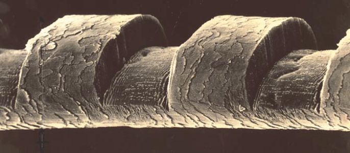
Fig. 1 – Scanning electron micrograph of a human hair etched by irradiation with an ArF
excimer laser; the notches are 50 ƒÝ wide.

While IBM Intellectual Property Law was preparing a patent application, we were constrained from discussing our discovery with people outside IBM. But we had a newly hired IBM colleague, Ralph Linsker, with an M.D. and a Ph. D. in physics. Linsker obtained fresh arterial tissue from a cadaver, and we irradiated a segment of aorta with both 193-nm light from the ArF excimer laser and 532-nm light from the Q-switched, frequency-doubled Nd:YAG laser. Once again, the morphology of the tissue adjacent to the irradiated/incised regions, examined by standard tissue pathology techniques, was stunningly different, with irradiation by the 193-nm light showing no evidence of thermal damage to the underlying and adjacent tissue.

This experimental study on freshly excised human tissue confirmed that excimer laser surgery removed tissue by a fundamentally new process. Our vision–that excimer laser surgery would allow tissue to be incised so cleanly that subsequent healing would not produce scar tissue–was more than plausible, it was likely, subject to experimental verification on live animals.

First public disclosure

After IBM filed our patent application on December 9, 1982, we were authorized to publicly disclose our discovery. We wrote a paper and submitted it to Science magazine, but it was rejected, because one of the referees argued that the irradiation of living humans and animals with far ultraviolet radiation would be carcinogenic, making our laser surgical technique more harmful than beneficial. Since Srinivasan now had an invitation to give a presentation about his work on polymers at the upcoming CLEO 1983 conference in Baltimore, MD, co-sponsored by the Optical Society of America, we wanted to get a publication into print as soon as possible, so we resubmitted our paper to the trade journal Laser Focus, including some remarks about the new experiments on human aorta. Serendipitously, the Laser Focus issue containing our paper was published simultaneously with CLEO 1983, where, on May 20, Srinivasan gave an invited talk entitled “Ablative photodecomposition of organic polymer films by far-UV excimer laser radiation.” In this talk, he gave the first oral public disclosure that the excimer laser cleanly ablated biological specimens as well as organic polymers.

From excimer laser surgery to ArF excimer laser-based refractive surgery

At this very same CLEO 1983 meeting, on May 18, Stephen Trokel and Francis L’Esperance, two renowned ophthalmologists, gave invited talks on applications of infrared lasers to ophthalmic surgery. I attended both of their talks and was amazed at the results they obtained in successfully treating two very different ophthalmic conditions. I was well aware that the ruby laser was first used to eradicate a retinal lesion in late 1961, and retinal surgery with lasers had become widespread in the ensuing two decades, in particular to repair retinal tears and to treat diabetic retinopathy. But these treatments required a laser at a wavelength for which the ocular media anterior to the retina was transparent. Excimer laser light would be absorbed in a thin layer upon entering the cornea, so the excimer laser would be useless for treating retinal maladies.

But Trokel knew of ophthalmic conditions, such as the refractive imperfection known as myopia, that could be corrected by modifying the corneal curvature. A treatment known as radial keratotomy (RK), developed in the Soviet Union and being practiced in the United States, corrected myopia by using a cold steel scalpel to make radial incisions at the periphery of the cornea. When these incisions healed, the curvature of the front surface of the cornea was reduced, with the consequence that the patient’s myopia was also reduced. The technique could rarely give the patient uncorrected visual acuity of 20/20, but the patient’s myopia was definitely reduced. One serious drawback of RK was that the depth of the radial incisions left the cornea mechanically less robust, and the healed eye was more susceptible to “fracture” under impact, such as might occur during an automobile collision. Trokel speculated that the excimer laser might be a better scalpel for creating the RK incisions.

Upon learning of our discover of excimer laser surgery, Trokel, who was affiliated with Columbia University’s Harkness Eye Center in New York City, contacted Srinivasan and subsequently brought enucleated calf eyes (derived from slaughter) to our IBM Research Center on July 20, 1983. Srinivasan’s technical assistant, Bodil Braren, participated in an experiment using the ArF excimer laser to precisely etch the corneal epithelial layer and stroma of these calf eyes. The published report of this study is routinely referred to by the ophthalmic community as the seminal paper in laser refractive surgery.

To conduct studies on live animals, the experiments were moved to Columbia’s laboratories. Such experiments were necessary to convince the medical community that living cornea etched by the ArF excimer laser does not form scar tissue at the newly created surface and the etched volume is not filled in by new growth. The first experiment on a live rabbit in November, 1983, showed excellent results in that, after a week of observation, the cornea was not only free from any scar tissue, but the depression had not filled in. Further histological examination of the etched surface at high magnification showed an interface free from detectable damage.

L’Esperance, also affiliated with Columbia’s Harkness Eye Center, thought beyond RK and, in November, 1983, filed a patent application describing the use of excimer laser ablation to modify the curvature of the cornea by selectively removing tissue from the front surface, not the periphery, of the cornea. His U.S. Patent No 4,665,913 specifically describes this process, which was later named photorefractive keratectomy (PRK).

Soon ophthalmologist around the world, who knew of the remarkable healing properties of the cornea, were at work exploring different ways to use to excimer laser to reshape the cornea.  From live animal experiments, they moved to enucleated human eyes, then to blind eyes of volunteers, where they could study the healing. Finally, in 1988, a sighted human was treated with PRK (Editors Note: by Dr. Marguerite McDonald at LSU), and after the cornea had healed by epithelialization, this patient’s myopia was corrected.

To read the complete article written by Dr. Wynne, including his footnotes, please follow this link.

AMD Update 25: Results of The AREDS2 HOME Study of Notal Vision’s Home Monitoring Device for AMD Announced

In April of 2010, I wrote about the inclusion of Notal Vision’s ForseeHome AMD Monitor in the AREDS2 clinical trial.
The overall objective of the two arm randomized clinical trial was to determine if home monitoring of participants at high risk of progression from late-stage dry AMD to neovascular AMD, using the comprehensive visual field and telemedicine solution based on the ForeseeHome Device in AREDS2 (referred to as the ForeseeHome comprehensive solution), would improve detection of progression to choroidal neovascularization (CNV) when compared with standard care (may have included use of the Amsler Grid).
Well, the results are in and the National Institute of Health (NIH) found that patients at high-risk for developing neovascular age-related macular degeneration would benefit from using the ForeseeHome Monitoring device for early detection of their CNV.
Report Represents the Most Comprehensive Study of Home Monitoring for Progression of AMD
As reported at the Retinal Subspecialty Meeting at this year’s AAO Meeting, the results of the Home Monitoring of the Eye (HOME) study, conducted in Age-Related Eye Disease Study 2 (AREDS2) clinical centers showed that participants at high risk for developing choroidal neovascularization (CNV) using the ForeseeHome monitoring device strategy had significantly better preservation of their visual acuity at the time of CNV detection than the control group of participants who were only using standard care methods (the Amsler grid) to self monitor their AMD for progression. The study’s Data Safety and Monitoring Committee recommended early termination of the study on April 2, 2013 based on superior vision outcomes among the participants randomly assigned to use the home device.
The AREDS2 HOME Study was a collaborative effort led by the National Eye Institute to evaluate the performance of a home monitoring device plus standard care compared to standard care monitoring alone for the detection of AMD progression to the neovascular phase. Standard care methods included periodic monocular self checks of vision clarity, blind spots and distortion, which included use of an Amsler grid. As treatments to manage the neovascular phase of AMD have improved, the importance of early detection of this event has increased in an effort to optimize outcomes following treatment of neovascular AMD. Approximately 8 million individuals in the United States, age 50 and older, are estimated to have intermediate (large drusen) or advanced dry AMD in one eye, placing them at high risk of progression to neovascular (wet) AMD (CNV), ranging from 25 to 50% over a five-year period.
Results of the HOME Study and Implications for AMD Management
At the time of CNV detection, 87% of eyes in the ForeseeHome device arm maintained visual acuity of 20/40 or better compared to 62% in the standard care alone arm. Median acuity among device users at the time of CNV diagnosis was 20/32. Among participants who used the device at the recommended minimum frequency (twice per week) to monitor their AMD for progression, 94% of eyes that progressed to CNV maintained 20/40 or better visual acuity. When CNV was detected, participants in the ForeseeHome device arm lost fewer letters on visual acuity testing (median loss of 4 letters) from entry levels of vision at the start of the study compared to those in the standard care alone arm (median loss of 9 letters). Use of the ForeseeHome device resulted in an increase in the proportion of CNV events first identified at home, meaning in between routine ophthalmic office visits to assess detection of disease progression. Among individuals using standard care methods for monitoring, only 55% of those that progressed noted symptoms at home that led them to present for examination; whereas 80% of the participants in the device monitoring group returned sooner than a scheduled visit because a change was noted by the device or by self-monitoring. This was associated with a greater degree of vision preservation at CNV diagnosis among individuals who returned promptly for changes, as the median visual acuity loss at CNV detection was 3.0 letters for those in the device arm compared with 11.5 letters for those in the standard care group. The average annual rate of false alerts among the device users, reported as the annual false positive rate, was 0.24 alerts/year, which may be extrapolated to one false alert on average every 4.2 years for each ForeseeHome user.
“Persons 60 years of age or older should undergo dilated eye examinations to determine their risk of developing advanced AMD, especially CNV,” said Jeffrey S. Heier, MD, Director of the Vitreoretinal Service and the Director of Retina Research at Ophthalmic Consultants of Boston and one of the principal investigators of the HOME Study. “In contrast to current home monitoring strategies, those with intermediate AMD (bilateral large drusen) or advanced AMD in 1 eye are likely to benefit from home monitoring with the ForeseeHome device to detect the development of CNV at an earlier stage with better preservation of their visual acuity to maximize visual acuity results after intravitreal therapy with anti-VEGF agents.”
About the HOME Study
The HOME Study was a controlled, randomized clinical trial that was part of AREDS2. The study was conducted in 44 clinical centers across the U.S., enrolling 1,520 participants at high risk for developing CNV. (With approximately half using the device and the other half acting as controls, using standard care.) The objective of the HOME Study was to determine whether monitoring with the ForeseeHome device plus standard care results in earlier detection of CNV compared to standard care alone. Standard care included instructions to the patient on self-monitoring for CNV. Better visual acuity at the time of CNV detection is both a reflection of earlier CNV detection as well as a favorable predictor for visual function outcomes following the management of CNV with intraocular anti-angiogenic medications.

About the ForeseeHome AMD Monitoring Program
The ForeseeHome AMD Monitoring Program is a prescription-based, comprehensive telemonitoring and data management system that extends the management of AMD to patients’ homes between office visits. The test results are transmitted to a central monitoring center that will alert, physicians to immediate, significant visual field changes in their patients, so that patients can be recalled for timely follow-up and necessary treatment may be initiated. The ForeseeHome AMD Monitoring Program utilizes a simple to use device based on preferential hyperacuity perimetry, a form of visual-field testing, to identify minute visual distortions, or metamorphopsia, for the detection of early CNV development.
To read more about Notal Vision and the ForseeHome device, read my full report of March 9, 2010: Notal Vision: The ForeseeHome AMD Monitor and It’s Potential to Save Vision – A First Report.

AMD Update 24: DARPins Phase 2 Trial Results Fall Short

Back in February, I first reported on Allergan’s DARPins in my Update 23: DARPins, The Next “Game Changer” for Wet AMD? In that report, I wrote that Molecular Partners’ MPO112 (Allergan’s AGN-150998) showed promise of improving vision and having a long ocular half-life which appeared to be a vast improvement over both Lucentis and Eylea, perhaps requiring injections every 3-4 months compared to bi-monthly for Eylea and monthly for Lucentis and Avastin. (I also noted a second agreement with Molecular Partners, the licensors of the DARPin technology to Allergan, in which a combination dual action anti-VEGF/PDGF drug therapy was also be under investigation.)
Well, the first part of the promise, the longer interval injection rate for the DARPins, has fallen through. As reported by two analyst groups, Allergan presented results last Friday (November 15th) from the Phase 2 trial of AGN-150998 (anti-VEGF DARPin program) in wet AMD at the Retina Subspecialty Meeting ahead of the start AAO annual conference in New Orleans. The results supported the company’s decision several months ago, to slow down advancement of the clinical trial, in that the drug failed to meaningful delay the time to retreatment and the associated rates of inflammation were higher than were anticipated. Though Allergan continues to evaluate the drug and still may ultimately advance it into Phase 3 studies, there appears to be only limited competitive threat to Eylea (or, perhaps Fovista, Ophthotech’s combination anti-VEGF/PDGF drug in clinical study – see my two write ups on Fovista, shown below, for more information about this potential drug). Specifically, the analysts see a low likelihood of commercial adoption or integration into the treatment paradigm for wet AMD without any sustained improvement in visual acuity or meaningful delay in the time to retreatment.
In looking at the data presented, the study evaluated two doses of the AGN-150998 (3mg and 4.2mg) vs. Lucentis. The drug was administered at week 4 and then pro re nata (PRN) or by week 16, and then again PRN or by week 32 at the latest. At day 60 and day 90, the 4.2mg dose appeared to delay the need for retreatment in ~10-15% of patients. Looking at the data another way, the 4.2mg dose appeared to delay the median time to retreatment by ~20 days. There were no differences in the percent of patients gaining 15 or more letters in best corrected visual acuity (BCVA) from baseline by week 16, and again at week 32.
In terms of safety, the AGN-150998 treatment was associated with a meaningful rate of ocular inflammation adverse events relative to Lucentis (13% vs. 0%). Specifically, treatment with AGN-150998 had higher rates of uveitis (3% with 3mg, 6% with 4.2mg, 0% with Lucentis), anterior chamber inflammation (2% and 3% vs. 0%), vitritis (7% and 2% vs. 0%).  For reference purposes, historical data imply the rate of intraocular inflammation in AMD trial are 13% and 1% with Lucentis and Eylea, respectively.
Allergan has indicated that it would be making changes to the manufacturing process to hopefully reduce the inflammation seen in the Phase 2 trials, when and if they decide to proceed to a Phase 3 trial.
I was not able to determine if Allergan and Molecular Partners still plan to go ahead with a clinical trial for the dual action drug, which remains in a pre-clinical stage.
References:
Analyst Reports – Private correspondence.
Fovista Reports:

Gene Therapy in Ophthalmology Update 21: New Gene Therapy Company, Spark Therapeutics, Launches

Children’s Hospital of Philadelphia (CHOP) announced that it had spun off its work in gene therapy to a new, fully integrated company, Spark Therapeutics, that will assume control over two current gene therapy clinical trials: a Phase III study for Leber’s Congenital Amaurosis, an inherited disease that results in blindness caused by mutations of the RPE65 gene, and a Phase I/II study for hemophilia B. The new company is also advancing toward the clinic with gene therapy programs to address neurodegenerative diseases and additional hematologic disorders and other forms of inherited blindness. One such program, in the latter category, already in pre-clinical development at CHOP, could be its study for the treatment of Choroideremia, a rare inherited disorder that causes progressive loss of vision due to degeneration of the choroid and retina.
Editors Note: It should be noted that one clinical trial using gene therapy to treat Choroideremia is already underway at Imperial College London and Oxford University, in conjunction with Moorfields Hospital in London.
The new company has been launched with a $50 million capital commitment from Children’s Hospital to advance and commercialize multiple ongoing programs with clinical proof of concept.
As noted by Susan Young, writing about the launch in Technology Review, “Spark has a chance to be the first gene-therapy company to obtain FDA approval. Results for a late-stage trial of a gene therapy for Leber’s Congenital Amaurosis … are expected by mid-2015. That treatment is one of several gene therapies in or nearing late-stage testing contending to be the first gene therapy approved by the FDA for sale in the U.S.”
The Phase III trial was initiated late last year, and CHOP has made significant progress in enrolling patients. Spark will be sharing additional details on its progress and encouraging results in the very near future.
And, as shown in my table of Ongoing Clinical Trials in Ophthalmology, it is the only gene therapy trial (in ophthalmology) that has advanced to Phase III.
Editors Note: For clarity, is should be noted that there are five other clinical trials underway to treat Leber’s, as shown in my table, but all are currently Phase I or Phase I/II studies. The Spark Therapeutics trial is the farthest advanced.
“The creation of Spark is the culmination of a decade-long commitment by CHOP and our founding team to drive the field of gene therapy forward during a time when many in the industry had moved away,” said Jeffrey D. Marrazzo, co-founder, president and chief executive officer of Spark Therapeutics. “Their vision and long-term dedication have enabled us to effectively address many of the key challenges facing the field and to emerge with one of the industry’s most robust clinical-stage gene therapy pipelines; as well as exclusive rights to commercialize a proprietary manufacturing platform, supply from a world-class manufacturing facility and a founding team with a proven track record of executing safe and effective gene therapy trials for nearly two decades. We are working with great urgency and care to deliver gene therapy products with the potential to transform the lives of those affected by severe genetic diseases.”
Spark builds on the work of CHOP’s Center for Cellular and Molecular Therapeutics (CCMT), established in 2004 as a world-class center for gene therapy translational research and manufacturing. Many of the CCMT’s leaders will assume management roles within Spark or engage with the company as scientific advisors, including Katherine A. High, M.D, a gene therapy pioneer who has served as the director of the CCMT since its inception.
“Gene-based medicines are among the most complex therapeutics ever developed,” said Dr. High. “We at CCMT have persevered through more than a decade of scientific and clinical development and are now closer than ever to realizing the ambitious vision of one-time, potentially curative therapies to address serious genetic conditions. The team at Spark has incredible goals for the treatment of diseases including hemophilia B and inherited blindness, and we look forward to working with them to deliver groundbreaking new treatments to patients in need.”
Spark has entered into agreements with multiple academic institutions to assemble the technology, programs and capabilities needed to deliver its pioneering gene therapy products. Notably, Spark has exclusive rights to commercialize CHOP’s proprietary manufacturing technology and will use clinical-grade gene therapy vectors produced by the CCMT’s state of the art good manufacturing practices (cGMP) clinical facility.

Pioneers in AAV delivery
Over the past two decades, the Spark leadership team has developed unrivaled expertise in the design, manufacturing and delivery of gene therapies using adeno-associated virus (AAV) vectors. AAV has been demonstrated in clinical studies to be a safe and effective vehicle for the delivery of genetic material into targeted cells and provides unique advantages over alternative delivery approaches. The Spark team was among the first to demonstrate human clinical proof of concept in two distinct organ systems — the eye and the liver — establishing a strong foundation for the company’s current programs, and has clinical experience in 15 studies across diverse genetic and non-genetic diseases and five distinct routes of administration.
Spark’s most advanced clinical program is a Phase III study to address blindness caused by mutations in the RPE65 gene. There is currently no pharmacologic treatment for this form of inherited retinal degeneration, which ultimately causes irreversible blindness.
The open-label, randomized, controlled study builds on an earlier clinical study in which 12 patients with RPE65-related blindness demonstrated notable improvement in visual function, moving in some cases from being profoundly blind to being able to recognize faces and ambulate independently. All school-age patients enrolled in the trial were able to transfer from Braille classrooms to sighted classrooms.
One such patient was Corey Haas, whose story is related in the book “The Forever Fix: Gene Therapy and the Boy Who Saved It”.
Corey Haas, his parents, and the CHOP team that treated his Leber’s and gave him back his vision.  
Read Corey’s story in Ricki Lewis’ book, The Forever Fix: Gene Therapy and the Boy Who Saved It.

The team’s experience in the clinical study of gene therapy – from designing and manufacturing vectors to conducting studies that have shown strong potential for safety and efficacy – is unparalleled in the field. Clinical-grade vectors prepared by the team have been used to safely treat more than 100 human subjects in 12 clinical trials in the U.S. and EU, across five parenteral routes of administration in genetic and non-genetic diseases. No other group can claim this breadth of expertise and experience in human gene therapy.
The adeno-associated virus (AAV) vectors used in the clinical programs have been demonstrated to be safe and effective vehicles for delivering genetic material into targeted cells, providing unique advantages over alternative therapeutic approaches. The team has established human proof of concept in two organ systems – the eye and the liver – and are advancing a Phase III program in blindness caused by mutations of the RPE65 gene; a Phase I/II program in hemophilia B; and preclinical programs in neurodegenerative diseases and other hematologic disorders and forms of inherited blindness.

Gene Therapy in Ophthalmology Update 20: Oxford BioMedica Clinical Trials Resume

Back in June, Oxford BioMedica announced that it had voluntarily paused recruitment for its clinical trials for wet AMD  (RetinoStat Phase I), Stargardt’s Disease (StarGen Phase I/IIa) and Usher’s Syndrome (UshStat Phase I/IIa). The company had halted recruitment of the aforementioned studies, as a precautionary measure, while it investigated the detection of very low concentrations of a potential impurity in its clinical trial material derived from a third party raw material.
Oxford has since performed extensive characterization studies using its newly developed, state-of-the-art analytical methods to identify the impurity as highly fragmented DNA derived from fetal bovine serum (FBS), the most widely-used growth supplement for cell culture media.  In light of these findings, Oxford remains convinced of the safety, integrity and quality of its LentiVector platform products and no safety concerns relating to any of the ocular products have been identified in any pre-clinical and clinical data generated to date.
Today, the company announced that following the submission of a comprehensive data package to the FDA and the French regulatory agency, ANSM, it has received agreement from both agencies to resume recruitment into its ocular clinical trials using the existing clinical trial material. The company will continue to use highly sensitive, state-of-the-art analytical methods to ensure the quality and integrity of its lentiviral vector products and will work with FDA and ANSM to define the necessary specifications for future batches of clinical trial material.
Oxford is now working closely with the clinical trial centers to obtain the necessary ethics committee approvals in order to resume recruitment into the clinical studies.
(For a list of the clinical site centers in the U.S. and France involved in the three studies, please take a look at my Gene Therapy Ongoing Clinical Trial Table at http://tinyurl.com/GeneTherapyClncal)
John Dawson, Chief Executive Officer of Oxford BioMedica, said: “We value our relationships with the regulatory authorities and are pleased that, on the basis of our extensive technical investigations to demonstrate the integrity of our products, FDA and ANSM agree with our proposal to resume treating patients in our ocular trials as soon as possible.
“We place the highest importance on safety, and our analytical methods and quality assurance processes are continuously evolving to ensure that we remain at the forefront of gene therapy development and manufacture. I am confident that, with significant opportunities ahead such as the recently-announced AMSCI project win, Oxford BioMedica will continue to lead the way in delivering novel gene therapies to patients.”
For your information, Oxford BioMedica has reported that 9 of the 18 patients to be treated in the wet AMD clinical trial had been treated; 12 of the 28 patients in the Stargardt’s trial; and 3 of 18 patients in the Usher Syndrome trial had been treated prior to the halt in recruitment in June.
Coincidently, Genzyme, who is also running a gene therapy clinical trial to treat the wet form of AMD, also announced a halt in recruitment for its trial in July. No reason for the stoppage has been given and all attempts to determine why the halt in recruitment occurred have been rebuffed. As of the last time I had obtained reliable information about the Genzyme trial, 6 of 34 patients to be treated had been treated.

Research in Retinal Disease: The Foundation Fighting Blindness Invests $2.1 Million in Seven New Research Efforts

As I continually search the web for interesting news about new technologies for treating retinal diseases, I came across this news from the Foundation Fighting Blindness’ website  yesterday afternoon. It relates to some of the annual grants to researchers that the FFB will be funding this year. It includes better ways of looking at retinal cells (via use of the adaptive optics laser scanning ophthalmoscope) and several projects involving gene therapy, along with a couple looking at ways of, hopefully, stopping the progression of dry AMD.

The following write up is reprinted with permission of the FFB.
August 15, 2013
The Foundation’s Scientific Advisory Board (SAB) recently completed its annual grants review process, leading to the allocation of $2.1 million in funding for seven new research projects, including those for identifying new disease-causing gene mutations, developing cross-cutting gene therapies and advancing potential treatments for dry age-related macular degeneration. The three-year grants were awarded after the SAB reviewed 117 proposals submitted to the Foundation last October.
“Grants review is a rigorous, multi-step process that takes most of the year to complete,” says Stephen Rose, Ph.D., chief research officer, Foundation Fighting Blindness. “Due to revenue limitations, we can only fund a fraction of the high-quality projects we’d like to fund. That makes the selection process even more challenging. We had to leave several excellent proposals on the table.”
Here are brief descriptions of the new research projects:
AOSLO: Detecting Retinal Degeneration Before Vision is Lost
The adaptive optics laser scanning ophthalmoscope (AOSLO) is like a powerful microscope that enables retinal researchers to see structural changes in the retina well before vision is lost from a retinal disease. That power can enable researchers to more quickly determine if a treatment is working in a clinical trial. Austin Roorda, Ph.D., of the University of California, Berkeley, is performing studies of AOSLO to correlate changes in the retina (e.g., loss of photoreceptors) with changes in vision.

Enhancing AOSLO for Expanded Clinical Use
Like Dr. Roorda, Stephen Burns, Ph.D., of the University of Indiana, is working with AOSLO to study the correlation between retinal and vision changes. He is also making AOSLO more affordable by using newer camera technology. In addition, he’s employing state-of-the-art computing technologies derived from video games to decrease image-processing times and costs. The new technology will make the imaging process more comfortable for the patient by tolerating more head and eye movement.
Figuring Out Why Severity of Vision Loss Varies for People with XLRP
Researchers have reported for many years that the severity of vision loss for people with X-linked retinitis pigmentosa (XLRP) can vary greatly, even for people within the same family. Stephen Daiger, Ph.D., of the University of Texas Health Science Center at Houston, will be looking at the role of a various biological, genetic and environmental factors in vision-loss variability for those with XLRP. The identification of a significant factor that modulates vision-loss severity – perhaps a protective protein – could lead to a potential treatment.
Finding New Genes Linked to ADRP
Researchers have identified almost two dozen genes linked to autosomal dominant retinitis pigmentosa (adRP), but many are yet to be found. Rui Chen, Ph.D., of Baylor College of Medicine, is on the hunt for those remaining adRP genes. With DNA from 118 adRP families, including 18 families with at least nine affected members, Dr. Chen is well positioned to identify additional genes linked to adRP. Finding the new genes will provide researchers with targets for treatments and cures.

Developing Neuroprotective Gene Therapies to Preserve Vision
John Ash, Ph.D., is developing gene therapies that have the potential to preserve vision in people affected by a broad range of retinal diseases. Unlike corrective gene therapies, which work only for conditions caused by a specific gene, Dr. Ash’s proposed treatments are designed to keep the retina healthy independent of the underlying disease-causing gene. He also believes the proteins delivered by his treatments – PIM-1 and STAT3 – will be less likely to cause damaging inflammatory side effects than some previously investigated neuroprotective proteins.
Targeting Inflammation to Halt AMD
Thanks to previous Foundation-funded genetic studies, researchers have strong evidence that the progression of age-related macular degeneration is associated with an over-active immune system. This ultimately leads to inflammation and cell death in the retinal pigment epithelium (RPE), a layer of cells that provides critical waste and nutritional support to photoreceptors. Loss of the RPE subsequently leads to loss of photoreceptors and vision. Jayakrishna Ambati, M.D., of the University of Kentucky, is developing a gene therapy that preserves the RPE by preventing the harmful sequence of immune-system events.
Boosting Cells’ Energy Supplies to Save Vision in AMD
Based on prior research, Deborah Ferrington, M.D., of the University of Minnesota, believes that mitochondrial dysfunction in the RPE plays a significant role in the development of AMD. Mitochondria are like miniature organs (organelles) within all cells that provide energy. When not working properly in retinal cells, they can lead to cell death and vision loss. Dr. Ferrington is evaluating compounds that help protect mitochondrial function in the RPE.

A New Approach to Treating Chronic Kidney Disease: Replenishing Your Nephrons

As some of you may know, I have chronic kidney disease (CKD). Thanks to my wife and the scare put into me by my nephrologist, that I would soon have to begin dialysis, I have managed to get my CKD  in remission, or at least under control. My GFR number (that indicates when you must start dialysis) has held steady, or actually gotten better since my wife put me on a strict diet and I have lost about 15 pounds – and my nephrologist took me off of lisinopril (for blood pressure control), which seemed to raise my GFR by about three points. So, it appears I won’t have to go on dialysis any time soon.
All that being said, and with my strong interest in the use of stem cells and gene therapy in treating retinal diseases in ophthalmology, I have been searching the web for research on the use of stem cells to treat kidney disease or to produce new nephrons, the kidney cells that filter the blood as it passes through the kidney – and that go bad or die causing CKD. I think I have found very early research of that possibility.
Earlier this week, I saw a news  release from the University of Queensland in Australia that said that Dr. Melissa Little and her research group at the Institute of Molecular Bioscience (IMB) have found a set of six genes that can prompt some types of adult kidney cells to regress to an earlier stage of development (stem cells) and act like the precursors to the cells of the nephron. Since it is death or damage of nephrons that causes chronic kidney disease, by forcing adult cells to act like early nephrons, they may have potentially found a way to trigger the growth of new filters in the kidney.
All of your nephron cells are formed before birth and people with fewer nephrons are at higher risk of kidney disease.
Note: Dr. Little is one of sources that I had found in my web search as she had published a paper on “Stem Cell Options for Kidney Disease” in 2008, and we had corresponded at the beginning of this year when I began my search for a stem cell answer.
“This discovery is the first of its kind and offers hope to patients with chronic kidney disease. If we can find a way to provide new nephrons to an adult or increase nephron numbers in babies at birth, we could potentially reduce the risk of disease progression,” said Professor Little.
This landmark paper, “Direct Transcriptional Reprogramming of Adult Cells to Embryonic Nephron Progenitors”, by Caroline E. Hendry, Jessica M. Vanslambrouck, Jessica Ineson, Norseha Suhaimi, Minoru Takasato, under the supervision of Professors Fiona Rae and Melissa H. Little, was published June 14th in the Journal of the American Society of Nephrology, the world’s leading nephrology journal.
Professor Little said, “There was still more work to be done to encourage these reprogrammed early nephron cells to function and integrate. While this is a beginning, we hope it will inspire industry leaders and researchers around the world to invest further in cellular and bioengineering approaches to kidney repair and regeneration.”
Stem Cells Australia Program Leader and Chair of Stem Cell Science at The University of Melbourne, Professor Martin Pera welcomed the research findings. “This innovative study provides evidence that adult cells can be reprogrammed to resemble the cells in the embryo that give rise to the kidney. The results pave the way for future studies that will enable researchers to produce human kidney cells in the laboratory, for use in studies of renal disorders, and for testing new drugs. Eventually this technology might help to make cells for transplantation to treat kidney disease,” said Professor Pera.
The Technology
I have attempted to read Dr. Little’s paper on reprogramming kidney cells, and with her assistance, this is what I understand she and her colleagues have done, which is a very early step in the long road to someday being able to replenish nephron cells in an adult kidney.
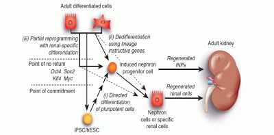
In an earlier paper written by Caroline Hendry and Dr. Little, “Reprogramming the kidney: a novel approach for regeneration”, they discussed the various approaches that might be taken to re-create viable cells within a diseased kidney, including using  induced pluripotent stem cells (iPSCs) derived from skin cells or other sites, or even the use of embryonic stem cells (ESCs) that would be introduced into the kidney to form new nephrons (?), if they could – as shown in the accompanying figure (but how would you control the formation of the new cells?). But they concluded that the best approach would be reprogramming existing kidney cells to the progenitor stage, with the hope that these would develop into the needed new cells, or in this case, nephrons, the approach they ultimately used in this new research.
Figure1 | The application of reprogramming to the kidney, indicating the feasible starting cells and target phenotypes.
(i) Reprogramming may involve the directed differentiation of human embryonic stem cells (hESCs)/induced pluripotent stem cells (iPSCs) to a renal lineage. The iPSCs may be recipient-derived and may be derived from adult kidney cells or any other available adult cell type using the same factors. Directed differentiation is likely to recapitulate development; hence, it is likely to require differentiation through a nephron progenitor intermediate (induced nephron progenitor cell; iNP) but may continue on to more specific mature renal cell types.
(ii) A specific renal lineage may also be achieved via lineage-instructive reprogramming directly to that state from an adult cell type. Again, this may be the renal epithelium, renal stroma, or any other available differentiated adult cell type; however, this is likely to be more successful if the attractor states of the starting and target cell type are as close as possible. Reprogramming may be to the iNP state or directly to a more mature renal cell fate.
(iii) Finally, reprogramming may use the classical Yamanaka factors until the cells pass the point of no return, after which a renal lineage may be reached via the application of the appropriate environmental cues. Such cues may once again target the iNP state or aim to directly induce a more mature renal cell type.
As previously stated, the nephron progenitor population of the embryonic kidney gives rise to all of the nephron cells that will be present in the adult kidney, prior to birth. So, currently, what you’ve got at birth is what you live with.
Using a screening technique, the researchers were able to identify a group of six genes, that activate a network of genes that can reprogram adult proximal tubule cells back to the nephron progenitor stage – which in turn can form adult nephron cells. Although the researchers believe that other factors are required, they concluded that these results suggest that re-initiation of kidney development (nephron cells) from a population of adult cells (proximal tubule cells) by generating embryonic progenitors may be feasible, opening the way for additional cellular and bioengineering approaches to renal repair and regeneration.
In their literature search, they could not find any previous reports of kidney cells being reprogrammed back to a progenitor cell type. Their hope is that this discovery will lead others to follow their lead and begin further work in the possible reprogramming of adult kidney cells for the repair and rejuvenation of diseased kidneys.
Sources:
News Releases:
Research reprograms future of kidney health, Institute for Molecular Bioscience and Stem Cells Australia, June 14, 2013
Papers:
Reprogramming the kidney: a novel approach for regeneration, Hendry and Little, Kidney International, March 21, 2012
Stem Cell Options for Kidney Disease, Hopkins et al, Jnl of Pathology, October 20, 2008

Gene Therapy in Ophthalmology Update 19: A New Virus Vector for Safer Delivery of Gene Therapies

Researchers at the University of California at Berkeley, along with some assistance from the Flaum Eye Institute and Center for Visual Science at the University of Rochester, have come up with a new version of an adeno-associated virus (AAV) vector that can deliver genes deep into the retina using an intravitreal injection of the vector into the vitreous, a less-invasive technique, instead of an intraretinal injection below the surface of the retina, which is the common way gene therapy is currently delivered.
The study was authored by postdoctoral fellows Deniz Dalkaral (then with Helen Wills Neuroscience Institute of UCal Berkeley, but now with Institut de la Vision in Paris) and Leah C. Byrne (Helen Wills), and graduate students Ryan R. Klimczak and Meike Visel (UCal Berkeley’s Dept. of Molecular and Cell Biology), and Lu Yin and William H. Merigan (Flaum Eye Institute and Center for Visual Science at the Univ. of Rochester), under the direction of Professors John G. Flannery, and David V. Schaffer of UCal Berkeley. The paper, “In Vivo–Directed Evolution of a New Adeno-Associated Virus for Therapeutic Outer Retinal Gene Delivery from the Vitreous”, was published online on June 12th in Science Tranlational Medicine.
As explained by Dr. Jean Bennett, a professor of ophthalmology at the University of Pennsylvania in Philadelphia, who was not involved in the study, but who has done extensive work with gene therapy in the treatment of Leber’s congenital amaurosis, “It shows the results of a very clever system to evolve AAV to target cells in the retina efficiently from an intravitreal injection.”
“Intravitreal injection, whereby a needle is pushed into the eye’s vitreous, or gel-like core, is a common drug delivery procedure performed under local anesthetic in a doctor’s office”, explained Bennett. “But using this routine injection technique in trials of gene therapy for retinal degeneration has thus far proven impossible.”
The problem, as explained by Dr. David Schaffer, a professor of chemical and biomolecular engineering, bioengineering, and neuroscience at the University of California, Berkeley, who led the research, is that current AAV vectors are incapable of penetrating deep into the retina where the target cells for retinal diseases are located. “AAV is a respiratory virus and so it evolved to infect lung epithelial cells,” explained Schaffer, “It never evolved to penetrate deep into tissue.”
Patients receiving gene therapy have theretofore undergone a vitrectomy (removal of the vitreous) and a direct intraretinal injection, which requires hospitalization and general anesthetic, and can sometimes even damage the retina. “If it were possible to inject AAV into the vitreous instead of the retina and still get gene delivery to the target cells, said Bennett, “one could envision the [doctor saying], ‘Ok, well just come into the office and get your gene therapy, tomorrow afternoon at two.’”
With that aim, Schaffer and colleagues used a process called “directed evolution” to randomly create millions of variations of the AAV virus to determine which ones were better at tissue penetration. They injected regular AAV into the vitreous of mouse eyes and one week later collected photoreceptor cells from deep within the retina. The tiny percentage of AAV vectors that made it into those cells were then amplified, repackaged into virus particles and injected into the vitreous again. They repeated the injection, recovery, and amplification a total of six times, finally isolating 48 AAV variants for sequencing. Two thirds of those isolates turned out to be the same variant, and Schaffer and colleagues named it 7m8.
Lastly, to determine whether the 7m8 vector would be likely to show similar deep penetration in the human retina, Schaffer had the vector fused to a fluorescent protein injected into the vitreous of macaque eyes. Primate retinas are considerably thicker than those of mice, and the vector did not consistently reach the deep cell layers – showing a spotty penetration pattern rather than the wide and even pan-retinal penetration that had been seen in the mice. However, 7m8 did effectively target photoreceptor cells of the fovea – a thinner part of the primate retina that is essential for the sharp detailed vision humans use when reading and driving. “That’s a really important region to protect,” said Schaffer. “For the quality of life of patients who are going blind, if you can at least protect the fovea that would be a huge improvement.”
Eye cells labeled with green fluorescent protein have successfully taken up the virus, showing that the ‘evolved’ virus (right) is more effective than the virus currently used for gene therapy (left). The new virus is particularly good at targeting the critical photoreceptors (top layer). (Source: University of California, Berkeley)
Note: All of the “directed evolution” work with mice to discover the 7m8 AAV vector was done at UCal Berkeley, while the confirmatory primate injection and imaging was done at the Univ. of Rochester.
Schaffer and colleagues don’t yet know what makes the 7m8 vector so much better at tissue penetration than its AAV ancestor, but they plan to find out and use that knowledge to further improve its penetration in the primate retina.
“Building upon 14 years of research, we have now created a virus that you just inject into the liquid vitreous humor inside the eye, and it delivers genes to a very difficult-to-reach population of delicate cells in a way that is surgically non-invasive and safe. “It’s a 15-minute procedure, and you can likely go home that day.”
The engineered virus works far better than current therapies in rodent models of two human degenerative eye diseases (X-linked retinoschisis and Leber’s), and can penetrate photoreceptor cells in monkeys’ eyes, which are similar to those of humans.
Schaffer said he and his team are now collaborating with physicians to identify the patients most likely to benefit from this gene delivery technique and, after some preclinical development, hope soon to head into clinical trials.
Schaffer predicts that the viruses can be used not only to insert genes that restore function to non-working genes, but can knock out genes or halt processes that are actively killing retinal cells, which may be the case in age-related macular degeneration.
As noted by  Dr. Stephen Rose, Ph.D., chief research officer, Foundation Fighting Blindness, one of the co-funders of the research, “This is a critical next step in the development of retinal gene therapies. The enhanced AAV holds potential for treating more of the retina and doing so more safely. Incremental advancements like this are essential to getting the best treatments out to the patients.”
The investigators showed efficacy for the 7m8 AAV in a large animal as well as mouse models of retinoschisis and Leber congenital amaurosis, or LCA (RPE65 mutations). In the mouse studies, the virus was able to penetrate the retina and deliver a corrective gene to enable the retina to function normally.
While the large animal did not have a retinal disease, the virus transduced many regions of its retina. Ultimately, in both types of animals, the AAV was able to deliver genetic cargo to a variety of retinal cells, including: photoreceptors, the cells that provide vision; the retinal pigment epithelium, a layer of cells providing nutrients and waste disposal; and ganglion cells, which are a target for emerging, vision-restoring optogenetic therapies. (Editor’s Note: See, for example, my writeup of the “Nirenberg Technique”, an optogenetic approach to restore near normal vision to the blind.)
Most notably, the intravitreally administered AAV was able to penetrate the fovea, a small pit in the center of the retina rich in cones, which provides the vision most critical to daily living, but is often made fragile by degenerative diseases. Researchers have been concerned that injections underneath the fovea could cause permanent damage and vision loss in patients with advanced degeneration in their central retina.
AAVs are currently used for gene delivery in several retinal gene therapy clinical trials, including those that have restored vision in children and young adults with LCA (RPE65). AAVs are attractive for gene delivery because of their natural ability to penetrate a variety of cells. In addition, humans are exposed to the virus in nature and, therefore, tolerate it well.
To identify the optimal AAV for intravitreal gene delivery, the scientists used a process called “directed evolution” to randomly create millions of variations of the virus. The variants were then screened in mice to identify the top candidates for gene delivery to the retina. In addition to looking for an AAV that could penetrate retinal cells well, the researchers searched for a variant that could pass through a formidable barrier in the eye known as the inner limiting membrane, or ILM, which separates the vitreous from the retina.
The scientists from UC Berkeley plan to perform additional toxicology and efficacy studies to ready the 7m8 AAV for study in humans.
Sources:

Researchers Identify Better Virus for Retinal Gene Delivery, Foundation Fighting Blindness, June 12, 2013

Stem Cells in Ophthalmology Update 25: ACT Patient in Dry AMD Trial Goes from 20/400 to 20/40!

The story started innocently enough. On Wednesday, May 15th, the journal Cell reported on a study that claimed biologists had finally created human stem cells by the same technique that produced Dolly the cloned sheep in 1996. They transplanted genetic material from an adult cell into an egg whose own DNA had been removed.
OK, an important story but what followed boggles the mind. Many science reporters wrote about the discovery which got picked up by several news sources. However, a sharp-eyed member of the Investor Stemcell Forum (iCell), a group originally started by investors in Advanced Cell Technology, noticed a quote at the end of Sharon Begley’s writeup. Sharon is a science reporter writing for Reuters. She had obtained quotes from several people working in the stem cell field, including at the very end of her story, the following quote from Dr. Robert Lanza, the Chief Scientific Officer of Advanced Cell Technology (ACT), “The most promising human study is ACT’s. It is two years into clinical trials using stem cells derived from human embryos to treat two forms of blindness, including macular degeneration, with encouraging results. One patient’s vision went from 20/400 to 20/40, said Lanza.
By Wednesday afternoon and into the evening, the iCell bulletin board went wild. There are more than 3000 members of this board and most are investors in ACT (or one of the other many stem cell companies discussed). Editor’s note: I am not a shareholder/investor, along with a few others, like Paul Knoepfler of Univ. of Cal. Davis, who are invited members because we write about the field. In the evening when I entered the board, there were several hundred comments all speculating whether the quote was accurate or, somehow, a mis-quote. This was the first time any of us had heard that a patient had achieved that level of correction.
Apparently, the SEC also took notice, because the next morning (Thursday, May 16th), the company was forced to acknowledge that the quote was accurate. In its statement, the company said, “Advanced Cell Technology today confirmed that the vision of a patient enrolled in a clinical investigation of the company’s retinal pigment epithelial (RPE) cells derived from human embryonic stem cells (hESCs) has improved from 20/400 to 20/40 following treatment. The improvement was first reported on May 15, 2013, in a news article published by Reuters.”

“We continue to be encouraged by the progress we see in our ongoing clinical investigations, though the results included in the article were confidential and not intended for publication at that time,” commented Gary Rabin, chairman and CEO of ACT. “Our plan is still to publish additional results from the clinical investigations when we have a significant aggregation of data.
Now we knew that a patient in one of the company’s clinical trials (there are three of them – two for treating Stargardt’s Disease – one in the U.S. and the other in the UK; and one for the dry form of AMD in the U.S.) had achieved unparalleled improvement. So, which clinical trial?
I speculated that it was probably in the dry AMD trial, because in the SMD trials, the patients treated had quite damaged photoreceptors which I didn’t believe could recover to that degree, but confirmation was still needed.
That evening, I asked the iCell board if anyone had confirmed which clinical trial had produced the significant results – and I got a private message providing the answer. I was right, it was a patient in the dry AMD clinical trial.
So, what is the significance of this development. It is significant because it shows that, for the first time, a person suffering with the dry form of AMD (90% of all those with AMD) can obtain improved vision, going from legally blind (20/400), to normal vision (20/40), good enough to obtain a driver’s license in most states. Yes, this is just one patient, and early in this clinical trial, but hope prevails. If this much improvement can be obtained with one patient, and that patient with very poor vision, than think what can be obtained starting with people with much less a degree of poor vision, 20/100, for example, which is part 2a of the Phase II clinical trial.
I am hopeful that ACT is on the right track.

A New Technique for Restoring Normal Vision to the Blind: The Technology of Prof. Sheila Nirenberg of Weill Cornell Medical School

About a year ago, a colleague put me in touch with a Boston-based venture capitalist who was interested in a new method for restoring vision to the blind that was under development at Cornell University. I did some cursory research about the technology and wrote a brief report about what I learned.

I really didn’t understand the front end of the technology – how the research team was able to acquire and manage a useful visual signal that could be converted into sight by the brain, but since the back end involved the use of gene therapy, which I was very interested in, I began a file to collect information about this technology.

Then, about a month ago, I learned that Dr. Sheila Nirenberg, the team leader, would be giving a presentation about her technology at an Optogenetics Conference that was being held in the Boston area. I contacted the symposium director and obtained an invitation to sit in on Dr. Nirenberg’s talk.

On May 1st, I went to the conference, met Dr. Nirenberg, listened to her presentation and literally got blown away!

I decided on the spot that I needed to learn more about what she and her team were doing and the best way was to write about it. So, I plunged in, did the research to learn more about what the Cornell team were doing, and with Dr. Nirenberg’s cooperation, here is my explanation of what I believe she and her team are doing and what her technology might be able to accomplish.
Introduction
Follow along with me for a minute. After a gene therapy injection (to place a light-activated dye into the ganglion cells of the retina) in a short procedure at a doctor’s office, a blind person suffering from retinitis pigmentosa, usher syndrome, or geographic atrophy (whose photoreceptors – the rods and cones – have been severely damaged) dons a unique pair of glasses (or goggles) and due to the magic of the Nirenberg conversion technology has his/her sight restored to something close to normal vision! That is what Dr. Nirenberg and her team is hoping to accomplish. She’s done it with animals so far and it raises exciting possibilities that it can be done with humans.
So, that raises these important questions: how does it work, how does it compare to other retinal prosthetic devices/methods being developed, and when will it become available for human clinical trials?
I will attempt to answer these questions.

How the Technology Works
In a normally-sighted person, the front of the eye focuses an image onto the retina. The image lands on the photoreceptors, which in turn sends the signals into the retinal circuitry, which processes them and converts them into a code. The code is in the form of  a series of electrical pulses, which get transmitted to the brain via the ganglion cells, which fine-tunes the visual information that is sent to the brain.
Fig. 1. The transformations of images into patterns of action potentials by the retina.
In a person with a retinal degenerative disease that destroys the photoreceptors, this train of events is short-circuited, and no light pulse information reaches the brain.  
What Dr. Nirenberg’s technology does is jump over the damaged tissue and contact the ganglion cells directly and drive them to send the code to the brain.       
The key to making this technology work, or the “eureka” moment, was when Dr. Nirenberg realized that what was needed was to provide a train of electrical pulses – the “code” – to the brain in a form that it was used to receiving and using to form an image or vision. In order to do this, she and her team came up with a two-fold approach, composed of an “encoder” to deliver the train of electrical pulses to the retinal structure that remained, and a “transducer” to recognize the “code” and transmit a similar pattern of electrical pulses to the visual cortex in the brain via the enhanced ganglion cells.
The encoder is composed of a pair of glasses or goggles that include a camera to capture what is being seen (think of the high-resolution camera in your iPhone or smartphone), a small programmable computer chip that converts the pixels seen by the camera into a coded pattern of electrical pulses that are “readable” or recognizable by the brain, and a mini-DLP (a mini-digital light projector) that transmits the light pulses to the retina (or that portion of the retina that contains the dye that can be activated by the light pulses).
I won’t get into how Dr. Nirenberg and her team came up with the algorithm that enables the procedure to work. That is aptly described in her paper (and the supporting information) recently published in Proceedings of the National Academy of Sciences (1).
The “transducer” portion, that enables the brain to “see” the train of light pulses, is composed of a light-sensitive dye (a protein – channelrhodopsin-2 or ChR2) that is injected into the eye, similar to the way Avastin is injected for treating the wet form of AMD, using a gene therapy technique called optogenetics, that places the dye into the ganglion cells of the retina.
As defined by Wikipedia, Optogenetics is a neuromodulation technique employed in behavioral neuroscience that uses a  combination of genetic and optical methods to control specific events in targeted cells of living tissue, even within freely moving mammals and other animals, with the temporal precision (millisecond-timescale) needed to keep pace with functioning intact biological systems.
In retinitis pigmentosa and other similar retinal diseases, the photoreceptors are destroyed, but the ganglion cells, which are part of the retinal system that are attached to the photoreceptors, usually are not, which is what makes them a prime target for vision restoration. By employing gene therapy to “carry” the light sensitive dye into the ganglion cells, the mini-DLP of the encoder fires the coded light pulses into individual ganglion cells, which in turn causes the light sensitive ChR2 dye to, in turn, fire light pulses which are carried by neurons via the visual cortex into the brain, which can recognize the code and turn it into vision.
In this way, what the camera in the glasses detect, is sent (in the proper form) to the brain which recognizes the signal and allows a blinded person to “see”.
Fig. 2. Images reconstructed from the blind retinas treated with the prosthetic. A. Original image. B. Left, image reconstructed from the firing patterns of the encoder. Right, image reconstructed from the firing patterns of the blind retina viewing the image through the encoder-ChR2 prosthetic. C. Image reconstructed from the firing patterns of the blind retina viewing the image through the standard optogenetic prosthetic (just ChR2, no encoder).

Competing Technologies – Retinal Prostheses, Stem Cells, Gene Therapy and Others Working with Optogenetics
As a means of putting Dr. Nirenberg’s technology into perspective, as one of the many approaches that are being developed for the ophthalmologist’s armamentarium for treating RP,  dry AMD, and other retinal degenerative disesases, I include the following information about the state of research for other experimental techniques/therapies that may find use in treating these diseases.
Retinal Prostheses (2)
In those with retinitis pigmentosa (RP) and similar retinal diseases, the retinal degeneration affects the retinal pigment epithelium and the photoreceptors. Eyes with RP respond to electrical stimulation because in many patients, the inner retina, particularly the ganglion cell layer, still has some function. The retinal chip implants stimulate these cells.
More than a dozen groups of investigators and companies around the world are working on retinal implants. In order to restore visual function, chip implants have to detect light, convert the light energy to electrical energy, and then stimulate the retina. Different groups approach this in different ways. Two of the implants that are furthest along the path to clinical availability are the Argus II Implant by Second Sight (now FDA approved), and the Active Subretinal Implant by Retinal Implant AG. The Argus Implant directly stimulates the ganglion cells. The Active Subretinal Implant recreates some of the signals that normally would have been made by the photoreceptors.
The Argus II Implant consists of four parts. The power comes from a battery pack worn on the hip. An external video camera wirelessly delivers images to the electrical housing that is affixed to the episclera. The image and data processing are done here. A cable from the electrical housing enters the eye through an incision in the pars plana and the electrical impulses then are sent through the cable to the chip. The chip itself is attached to the retina with a tack.
In clinical testing of the Second Sight implant, all 30 patients who received the implant during the trial were able to perceive light during stimulation. More than half of the patients were able to see the motion of a white bar moving across a black background. Many of the implanted patients were able to identify some 3 to 4.5 cm letters on a high-contrast background. The best vision to date was 20/1,262.
The Active Subretinal Implant is currently in clinical trials in several European centers and in Asia. This implant contains a 1,500-electrode array that directly stimulates the inner retina. In contrast to the Argus II implant, which bypasses the inner retina, the Active Subretinal Implant aims to replace the dysfunctional photoreceptors.
The Active Subretinal Implant contains photodiodes on the subretinal chip, so there is no camera. The light stimulation occurs similar to the way we see-the light coming from an object goes through the pupil and activates the implant, which then converts the light directly into electrical stimulation. In contrast to the epiretinal implant, the subretinal implant does the image processing within the chip itself. However, using this technology requires more energy than light can provide. This is provided via a handheld battery pack that also has controls for brightness and contrast. The necessary energy is transmitted transdermally via a receiver induction coil and a magnet that is implanted under the skin behind the ear. A subdermal cable tacked to the lateral canthus connects the receiver to the subretinal implant for energy.
There are published reports on a total of 21 patients who have received the subretinal 1,500-photodiode implant. Those patients have achieved VA of up to 20/1,000 within an 11 degree by 11 degree visual field. Functional outcomes included localization of objects of daily life such as plates and drinking glasses; increased mobility; motion detection; orientation in outside environments; recognition of facial details; even reading and detecting spelling errors in words written in letters 6-8 cm in size.
The basic take-away from a review of the work being done with direct retinal implants is that they are limited by the number of photodiodes that can be implanted – and by the direct light pulse information that can be transferred to the implant. The key, as I see it, is that Dr. Nirenberg has invented a “better” way to input a visual signal and unless that is incorporated into the retinal implant systems (as she has proposed to with Second Sight), they will never approach the conversion rate that she claims to have achieved.
Stem Cells, Cell (Drug) Therapy, and Laser Treatment
As I have previously written (3), a number of companies/institutions are using both adult and embryonic stem cells to invigorate the retinal epithelial layer that feeds the photoreceptors, in the hope of regenerating some activity in the photoreceptors. One company, Neurotech, is using encapsulated human RPE cells to secrete ciliary neurotrophic factor CNTF), which they believe is capable of rescuing and protecting dying photoreceptor cells.
Meanwhile, Ellex Laser has a research program aimed at “retinal regeneration” by using its laser to stimulate the RPE cells to release enzymes that are capable of “cleansing” Bruch’s membrane in the hope of rejuvinating the retina (photoreceptors) by allowing the increased transport of water and chemicals across this important membrance. (This technique might have some bearing in macular edema and in the early stages of dry AMD in drusen reduction, but I don’t see how it would affect the photorecptors (4).)

Gene Therapy and Optogenetics
As is clearly pointed out in my table on the use of gene therapy in ophthalmology (5), a number of companies and institutions are in the pre-clinical and clinical stages of developing gene therapy approaches for the treatment of dry AMD (geographic atrophy), RP and Usher’s Syndrome. Hemera and Oxford BioMedica are taking straight gene therapy approaches, while a number of companies/institutions are involved in using optogenetic gene therapy. Among those using optogenetics that I am aware of, are EOS Neuroscience, GenSight Biologics, RetroSense, the University of California at Berkeley, and the Instituite de la Vision (Paris).
Of course, it should be mentioned that Dr. Nirenberg is working with Dr. William Hauswirth of the University of Florida, in her pursuit of an optogentic approach to solving the problem of restoring vision for the blind.
Again, as I noted in the conclusion to the section on retinal implants, my belief is that none of these techniques (except of course for the work of Dr. Nirenberg and Hauswirth) should accomplish as good results without the inventive front end visual signal supplied by Dr. Nirenberg’s work. Input equals output and the best input should provide the best output!
Status of the Invention
As reported to me by Dr. Nirenberg, the original work done with mice has now led to work with primates. Her lab has constructed a device for use with the primates and, in conjunction with Dr. Hauswirth, they are now testing an array of channelrhodopsin-expressing vectors to be able to select the best candidates for a Phase I/II human clinical trial. As she states (6), “For a vector to serve this purpose, it has to a) produce normal firing patterns in blind retinas (as has been done in the mouse), and b) produce normal firing patterns in the specific cell classes we target, which are ganglion cells or subclasses of ganglion cells.”
“We are currently working with 8 AAV-2 vectors; they vary in the channelrhodopsin used, in the promoter to drive expression, and in the enhancer components. So far, at least 2 of the vectors satisfy these conditions, that is, they express channelrohodopsin in primate ganglion cells in vivo, and they express it strongly enough to allow normal firing patterns to be produced when they’re stimulated by the device. The next steps are to modify the vectors for humans and to perform safety studies (typically, studies in two species are recquired), and then (to) prepare a package for FDA for evaluation.”
“Thus, although it is likely that there will be hurdles to overcome to bring this technology to patients, the major ones – a vector (AAV) for delivering channelrhodopsin to ganglion cells, and encoder/stimulator device to drive them, and the fact that targeting a single ganglion cells class by itself can bring substantial vision restoration – have already been addressed, substantially increasing the probability of success.”
In conclusion, I should note that with all of the previous work done in the 16 gene therapy in ophthalmology clinical trials either currently underway or completed, the time to get into a clinical trial with the specific gene therapy vector chosen, should be short, rather than long. And then, the real test to demonstrate the ability of the Nirenberg Technique to restore vision to the blind, will begin.
Note: To view a presentation similar to the one I sat in on on May 1st,  please take a look at Dr. Nirenberg’s presentation at TED MED 2011 in October 2011.
Footnotes:
1. Retinal prosthetic strategy with the capacity to restore normal vision, Sheila Nirenberg and Chethan Pandarinath, PNAS, August 2012; Supporting Information, same source.
2. Retinal Prostheses Offer Hope to Blind Patients, Sunir Garg, MD, Review of Ophthalmology, March 15, 2013
3. A Primer on the Use of Stem Cells in Ophthalmology, Irv Arons’ Journal, Sept. 2010
6. Research Interests, Sheila Nirenberg, March 13, 2013.

Recently Published Articles: Current Status of Stem Cells and Gene Therapy in Ophthalmology

In the past couple of months, I was asked to update an article I wrote on stem cells in ophthalmology, originally published in Retina Today, for its sister publication, Advanced Ocular Care, and to write a similar article about the current status of gene therapy for another ophthalmic publication, Retinal Physician. These two articles have now been published in the respective journals and made available online.
Here is a brief summary of each article, along with the link to its online version and a note about finding the current versions of the tables associated with each, online.
The Current Status of Stem Cells in Eye Care, Advanced Ocular Care, March 2013
As noted, this is an update of the original article that appeared in the May/June issue of Retina Today.
“From an inauspicious start several years ago, the use of stem cells in the treatment of several ocular and retinal diseases has picked up steam over the past year.”
The article goes on to describe what stem cells are, the applications of stem cells in the various parts of the eye, a brief discussion of the status of some of the clinical trials, and concludes with a quote from Dr. Stephen Rose, chief research officer of the Foundation Fighting Blindness, who wrote, “Of course, it would be nice if all parts of our bodies, including our retinas, came with extended warranties so you could just swap them out when they go bad. But now that I think about it, that’s what stem cells might do for us someday.”
“With the first approval of a gene therapy treatment for treating a genetic disorder in the Western world, the future of gene therapy for treating ocular disorders looks bright.”
The article goes on to discuss what gene therapy is and how it works; the applications of gene therapy in ophthalmology and clinical trial status for four ocular diseases – Leber’s Congenital Amaurosis, wet AMD, Stargardt Disease, and Usher Syndrom 1b; attempts to answer some remaining questions; and concludes with a quote from officials with the Office of Cellular, Tissue and Gene Therapies (OCTGT) for the FDA, “The recent history of gene therapy has been a mixture of promise and disappointment … Despite the setbacks of the past, the OCTGT shares the enthusiasm of the field and is confident that ongoing clinical investigations will lead to commercially available gene therapy products that are safe and effective and advance the public health.”
Current Versions of Stem Cells and Gene Therapy Tables
Because of the lag between submission and publication of the above articles, the tables that are linked to the print and online versions of the above articles are currently out-of-date. I constantly update their contents and publish the latest versions online, which are accessible from my blog entry about each set of tables:
Stem Cell/Cell Therapy Companies/Institutions Active in Ophthalmology
A list of thirty-two companies and institutions working with stem cells/cell therapies for ophthalmic applications. The table lists collaborators, the cell type being used, and the applications for which the cells will be used.
Stem Cell/Cell Therapy in Ophthalmology by Application
A list of sixteen ophthalmic applications being studied in clinical trials. The table includes the companies/institutions involved, the clinical trial status, and an active link for the clinical trial for those listed. (Thirty-six active and completed clinical trials are shown.)
Stem Cell/Cell Therapy in Ophthalmology — Ongoing & Completed Clinical Trial Details
A list of the the sixteen ophthalmic applications and the thirty-six clinical trials showing the number of patients to be studied in each trial and the number studied to date (that I am aware of). Active links are provided for each ongoing or completed trial.
Gene Therapy Companies/Institutions Active in Ophthalmology
The table lists more than thirty-two companies and institutions actively pursuing gene therapy solutions to ophthalmic diseases. The table shows the delivery viral platform, the gene type being used (where known), the application, and clinical status.
Gene Therapy in Ophthalmology by Application
This table, like the one for stem cells, lists the nineteen ophthalmic indications, the company/institutions involved, the clinical status, and the clinical trial number. (Sixteen active clinical trials are listed, with live links.)
Gene Therapy in Ophthalmology — Ongoing Clinical Trial Details
Again, as with the stem cell clinical trial table, this table lists the sixteen active and completed clinical trials, the number of patients to be treated and the number of patients treated to date.
Note: The links in the tables associated with the clinical trials are “active” or “live” and will take you to each clinical trial on CinicalTrials.gov

A Personal Journey: How I Went From Being A Bench Chemist to An Expert Resource in Ophthalmology and Medical Lasers

A short while ago, I was asked by Maureen Duffy, editor of VisionAware, the blog of the American Foundation for the Blind, how I became so knowledgeable about ophthalmology and why I started my blog. I prepared some background information for Maureen and she published it as a guest blog on her site, but because of space limitations, she was only able to use an abridged version. Since I don’t have the same space limitations, I decided to publish the “unabridged” version here.

So, here is my story:

The Beginning of My Career in Chemistry
I graduated from the Univ. of Massachusetts in Amherst, MA in 1957 with a B.S. degree in organic chemistry. After three jobs in industry (The Refinery Technology Laboratory of Gulf Oil in Philadelphia – analyzing oils and gas products from the refinery; the Container & Sealant Lab at the Dewey & Almy Div. of WRGrace in Cambridge, MA – working on cap and can sealants for baby food and peanut butter jars and aerosol can valves; and The Exploratory Development Lab at United-Carr Inc. in Watertown, MA – working on adhesives to replace fasteners for automotive applications), I joined the staff of the Product Technology Section of Arthur D. Little (ADL) of Cambridge, MA, the international consulting firm, in March 1969, as a laboratory bench chemist.
Over my twenty-five years at ADL, I worked on hundreds of projects. Among the more memorial assignments were being part of the team that developed the “all plastic pencil” for the Empire Pencil Company (part of Hasbro Toy); working on the development of an erasable pen ink compound for Bic Pen; trying to produce a protective plastic liner for glass Thermos bottles (to protect the food contents of the bottles when school children hit their neighbors on the head with their lunch boxes – which proved a technical success – water, soup and coffee stayed hot overnight, but failed miserably, as the plastic liner took the flavor out of the coffee making it taste terrible!); developing a spin-cast epoxy eyeglass frame compound and manufacturing process for Universal Optical; developing an improved firefighter’s glove for NIOSH and NASA; the development of a unique, disposable, no moving-parts mixer for use in dispensing two-part epoxy and urethane adhesives for the MPB Corporation (now commonly used in all dentist’s offices for dispensing casting compounds and sold in hardware stores for delivering reactive adhesives and sealants); and of course being a part of the team that built and flew a lead balloon to prove that lead balloons really could fly! (In fact I tried to launch the silk purse made from sow’s ears in a basket beneath our lead balloon!)
I have written the stories behind many of the above inventions and products in my “other” blog, ADL Chronicles.
An Introduction to Soft Contact Lenses
One of my first assignments at ADL (along with working on chemically stabilizing soils for soft ground tunneling) was to see if there were other uses for hydroxy ethyl methacralate (HEMA), the material that soft contact lenses were composed of. National Patent Development Corp. (NPDC), which had acquired the Czech technology and licensed it to Baush & Lomb, wanted to know if this product could be used for other applications. We thought of applying it to the hull of boats to make them more slippery (and go faster in water and reduce adhesion of barnicles), but the best I could come up with was its use as a slow release reservoir for insecticides to kill mosquito larvae in swampy areas.
A few years later, in June 1972, and because of my knowledge of the properties of HEMA, NPDC asked me to lead a study on the “safety and efficacy” of the B&L Soflens, which had just been approved for marketing (March 1972). (NPDC was going to issue some stock to the public and the underwriters needed this due diligence report for the offering.) The study involved interviewing all of the original FDA clinical investigators for the Soflens. The knowledge about soft lenses I gained led to several papers about the “Outlook for Soft Lenses” that were published by ADL’s Decision Resource Division. Those publications led, over the next fifteen years, to over 150 assignments in the soft lens industry, including about 50 briefings to companies interested in both the technology and financial aspects of soft contact lenses. (How was B&L making so much money with their Soflens business?) It also led to my assisting Johnson&Johnson (Vistakon), Ciba Geigy (CibaVision), and Schering Plough (Wesley-Jessen) in their acquisitions of technology or small companies, to enter the soft lens industry. (In essence, I became the “guru” of the soft contact lens industry!)
Ophthalmology, Lasers and Writing
The soft lens assignments led to assessments of intraocular lenses (IOLs), plastic eyeglass lenses and eventually, in 1983, the just FDA-approved YAG ophthalmic lasers for correcting capsule opacities following cataract procedures. In 1985, I became involved with surgical lasers and in 1986 wrote the first comprehensive research and market report on medical lasers, “Medical Laser Systems: The Surgical Revolution”, that was published by Arthur D. Little’s Decision Resources Division. This was followed by a second comprehensive report on medical lasers, “The Outlook for Medical Lasers: The New Technologies”, published in 1988, also by Decision Resources. That led to my second career at ADL,  involving ophthalmic and medical laser consulting.
In the summer of 1985 I attended a Gordon Research Conference on lasers used with biological materials, where I met many of the leading researchers and surgeons involved in ophthalmic and medical laser technology. In December 1985, I was asked to prepare a market report about the potential for the excimer laser to correct vision. That report, about what I called LRK (Laser Photorefractive Keratectomy), became PRK, or ablation of the surface of the cornea to correct vision. My report was used to raise funds to begin one of the first excimer laser companies, Tauton Technologies (which later acquired VISX and assumed the VISX name).
That led to my entry into consulting in refractive lasers and writing the first market reports about refractive surgery in the mid-1980s and early 1990s. (I later wrote the seminal report on the future of laser refractive surgery for Summit Technoloy in 1992, wherein I predicted that the excimer laser would gain FDA marketing approval would occur by mid-1995. As I recall, I believe I was off by about 4 months.)
As I had begun attending ophthalmic and medical laser industry meetings (AAO, ASCRS, ARVO, ASLMS), I started writing about what I learned at those meetings. I first began writing columns for Vision Monday in 1988, mostly about my contact lens-related work. In 1989, I was asked to write a monthly column for Ophthalmology Management which became my Technology Update column. (Ophthalmology Management stopped publishing in the spring of 1991.) In the fall of 1991, Ocular Surgery News asked me to continue my Technology Update columns for them. This monthly feature, mostly covering new technologies that I discovered at the ophthalmic meetings, ran for over eleven year in OSN.
I was among the first to write about “custom ablation” and LASIK. One of my favorite titles from those times was, “Inlays, Onlays, Rings & Things” which described alternatives to laser refractive surgery.

Retirement and Spectrum Consulting
Finally, in March of 1994, on the anniversary of my 25th year with ADL, I retired from ADL and started my own consulting firm, Spectrum Consulting, continuing my consulting work in ophthalmics and medical lasers. In the fall of that year I started publishing Executive Laser Briefing, a monthly newsletter about current developments in the ophthalmic and medical laser industries. It was begun as an added service for my consulting clients, but I soon realized that others were interested in the publication. I began marketing the newsletter, which grew to a 40-60 page monthly publication, sent out to clients around the world, and it became a major part of my consulting business.
In December 2005, after eleven years, I  sold the newsletter and its client list to the publishers of Trends-In-Medicine, who continue to publish the newsletter today, renamed Executive Laser Report, and retired from active consulting.

A New Blogging Career Begins: Irv Arons’ Journal
That’s when I decided to begin writing my blog, Irv Arons’ Journal. It was originally started as a vehicle to place the more than 150 previously published articles online and accessible for historians and researchers, as I was involved in the beginning of several ophthalmic firsts – among them the birth of soft lenses, IOLs, ophthalmic lasers, refractive lasers, etc. Most of the articles were not available online because they were written prior to the explosion and availability of the web.
However, I quickly became diverted because I realized that I had insight into several new technologies that were entering ophthalmic use and I had access to those in the know who could provide me with the insight to write about these developments.
It all began because of some colleagues writing about what had happened the previous summer at the Retina 2005 Meeting in Montreal. Dr. Phil Rosenfeld presented information about his use of Avastin to stop the wet form of age-related macular degeneration (AMD) cold in its tracks and to provide improved vision to some patients. His presentation wowed the audience, but other than the retinal surgeons present at the meeting, no one else in the ophthalmic world was aware of the significance. I decided I needed to tell this story – Avastin: A New Hope for Treating AMD — and so began my blogging career with an emphasis on new technologies for treating retinal diseases.
Since that start in December 2005, I now have over 270 articles posted online, mostly about new treatments for both dry and wet AMD, but also including writeups about new devices used to both treat and detect AMD.
Along the way, I have also written about the use of lasers to treat eye floaters, about the history and use of femtosecond lasers, including their use in treating cataracts, an overview of treatments for glaucoma, including the use of the then new SLT laser.

A Developing Interest in Stem Cells and Gene Therapy
Over the past several years, I became interested in the use, first of stem cells, and then gene therapy in the treatment of ophthalmic diseases. In September 2010 I wrote A Primer on the Use of Stem Cells in Ophthalmology, which was followed in November 2010 with my first writeup on the use of gene therapy in ophthalmology, The Use of Gene Therapy in Treating Retinitis Pigmentosa and Dry AMD. Since then, I have followed with more than 15 updates on stem cell treatments and more than a dozen for gene therapies.
In addition, I have put together comprehensive tables of the companies and institutions involved in both stem cells and gene therapies, the ophthalmic applications under evaluation, and data about the clinical trials underway and completed for both therapies. All of this information is now available online via my blog.
And, the Story Continues
I hope this overview has given you a glimpse into how I went from being a bench chemist to becoming an authoritative resource in the new technologies for treating ophthalmic diseases and conditions, starting at the front of the eye and working my way into the back of the eye.
For continual updates, please visit Irv Arons’ Journal and follow me on Twitter @iarons.

People in the NorthWest with X-linked retinoschisis

XLRS Natural History Study Beginning in Portland, Oregon

March 29, 2013 – Oregon Health & Science University (OHSU) is launching a three-year natural history study for people with X-linked retinoschisis (XLRS). Funded by the Foundation Fighting Blindness, the investigation’s primary goal is to identify outcome measures — such as changes in vision or retinal structure — that could be useful in evaluating the effectiveness of potential therapies in clinical trials. The study will also help determine the types of XLRS patients most suitable for future therapeutic studies.

Knowledge gained from the XLRS natural history study will aid in the design of an XLRS gene therapy clinical trial slated to begin in late 2014 or early 2015. The trial will be a collaboration between Applied Genetic Technologies Corporation, OHSU and the Foundation.

X-linked retinoschisis occurs almost exclusively in males. Participants in the natural history study must be of that gender. Otherwise, to qualify, they must:

  • have a clinical diagnosis of XLRS
  • have a disease-causing mutation in the gene RS1
  • be 7 years of age or older
  • be able to provide consent/assent (understand study procedures and risks)
Participants will need to make yearly visits to the Casey Eye Institute in Portland, Oregon. Some travel may be reimbursed.
If study participants are not already using carbonic anhydrase inhibitors (CAI), they will be offered this standard-of-care treatment during the study. If participants start CAI treatment during the study, they will need to travel to OHSU for some additional visits. CAIs are thought to reduce retinal edema (swelling) and other symptoms associated with XLRS.
For more information about the XLRS natural history study, contact the study coordinator at (503) 494-2363.

Gene Therapy in Ophthalmology Update 18: A RetroSense Update

I first learned about the potential of using gene therapies in treating ophthalmic disorders back in November 2010. That’s when I was introduced to gene therapy by Sean Ainsworth, the founder and CEO of RetroSense Therapeutics. I haven’t written about this company or the unique approach it is taking to try and treat retinitis pigmentosa and the dry form of AMD since that first article, The Use of Gene Therapy in Treating Retinitis Pigmentosa and Dry AMD. With several news events occurring with the company recently, I felt it was time to bring readers of this blog up-to-date.
First, a brief review of the approach that RetroSense is taking. The technology that the company is using was developed at Wayne State University by Dr. Zhuo-Hua Pan. It involves using channelrhodopsin-2, delivered via an adeno-associated viral vector (AAV) directly into the retina to restore lost vision. Channelrhodopsin-2 is an “opsin”, derived from green algae which can be used to convert light-sensitive inner retinal neurons into photoreceptor cells, thereby imparting light sensitivity to retinas that lack photoreceptors. This is a process called “optogenetic therapy”.
As reported in Retina Today(1), “We took a new strategy for restoring vision by genetically converting the retina’s second- or third-order cells to become light sensitive to mimic the function of rods and cones,” wrote Dr. Pan. “But critical to this strategy, we needed to find certain suitable light sensors that can be easily inserted into these surviving retinal cells.”
Optogenetics is defined(2) as, “…the combination of genetic and optical methods to control specific events in targeted cells of living tissue, even within freely moving mammals and other animals, with the temporal precision (millisecond-timescale) needed to keep pace with functioning intact biological systems.”
           
In an interesting write-up about both optogenetics and RetroSense, Susan Young writing for MIT’s Technology Review(3), said, “The idea behind Retrosense’s experimental therapy is to use optogenetics to treat patients who have lost their vision due to retinal degenerative diseases such as retinitis pigmentosa. Patients with retinitis pigmentosa experience progressive and irreversible vision loss because the rods and cones of their eyes die due to an inherited condition.”
She went on to say, “Retrosense is developing a treatment in which other cells in the retina could take the place of the rods and cones, cells which convert light into electrical signals. The company is targeting a group of neurons in the eye called ganglion cells. Normally, ganglion cells don’t respond to light. Instead, they act as a conduit for electrical information sent from the retina’s rods and cones. The ganglion cells then transmit visual information directly to the brain.”
“Doctors would inject a non-disease causing virus into a patient’s eye. The virus would carry the genetic information needed to produce the light-sensitive channel proteins in the ganglion cells. Normally, rods, cones, and other cells translate light information into a code of neuron-firing patterns that is then transmitted via the ganglion cells into the brain. Since Retrosense’s therapy would bypass that information processing, it may require the brain to learn how to interpret the signals.”
Before I relate the latest news about the company, I would like to share one further write-up about the company’s technology. This brief appeared in February, as part of an article in Popular Science entitled, “How Neuroscience Will Fight Five Age-Old Afflictions(4)”. One of the “afflictions” noted was blindness, and the writeup described RetroSenses’ approach to curing that affliction.
BLINDNESS

Gene therapy converts cells into photoreceptors, restoring eyesight

Millions of people lose their eyesight when disease damages the photoreceptor cells in their retinas. These cells, called rods and cones, play a pivotal role in vision: They convert incoming light into electrical impulses that the brain interprets as an image.

In recent years, a handful of companies have developed electrode-array implants that bypass the damaged cells. A microprocessor translates information from a video camera into electric pulses that stimulate the retina; as a result, blind subjects in clinical trials have been able to distinguish objects and even read very large type. But the implanted arrays have one big drawback: They stimulate only a small number of retinal cells—about 60 out of 100,000—which ultimately limits a person’s visual resolution.

A gene therapy being developed by Michigan-based RetroSense could replace thousands of damaged retinal cells. The company’s technology targets the layer of the retina containing ganglion cells. Normally, ganglion cells transmit the electric signal from the rods and cones to the brain. But RetroSense inserts a gene that makes the ganglion cells sensitive to light; they take over the job of the photoreceptors. So far, scientists have successfully tested the technology on rodents and monkeys. In rat studies, the gene therapy allowed the animals to see well enough to detect the edge of a platform as they neared it.

The company plans to launch the first clinical trial of the technology next year, with nine subjects blinded by a disease called retinitis pigmentosa. Unlike the surgeries to implant electrode arrays, the procedure to inject gene therapy will take just minutes and requires only local anesthesia. “The visual signal that comes from the ganglion cells may not be encoded in exactly the fashion that they’re used to,” says Peter Francis, chief medical officer of RetroSense. “But what is likely to happen is that their brain is going to adapt.”

Rewiring The Brain: Blindness: a) An eye diseased with retinitis pigmentosa has damaged photoreceptors, or rods and cones. Doctors inject the eye with a nonharmful virus containing the gene channelrhodopsin-2, or ChR2. b) The virus migrates into the retina at the back of the eye and inserts the gene into ganglion cells, which relay signals from the rods and cones to the optic nerve. The ganglion cells begin expressing the ChR2 protein in their membranes. c) Incoming light activates the ChR2 protein in ganglion cells, stimulating them to fire an electrical impulse. That message travels through the optic nerve to the brain’s visual cortex, which interprets it as a rough image.  Medi-Mation (Used courtesy of Popular Science)
What’s New
Within the past few weeks, the company has made two important announcements relative to its intellectual properties:
On March 5th, the company announced the notice of allowance for a new U.S. Patent Application broadly covering optogenetic approaches to vision restoration. The Patent Application broadly covers methods of restoring visual responses with a variety of optogenetic compounds. Specifically, the allowed application includes claims covering methods of restoring visual responses by delivering channelrhodopsin and variants thereof, as well as halorhodopsin to retinal neurons – with or without the use of cell-type specific promoters, including mGluR6 (Grm6). The subject opsins have been studied extensively and published on as means of vision restoration in retinal degenerative conditions such as retinitis pigmentosa and dry age-related macular degeneration.
The approved patent application is part of the “Pan” patent family, which stems from the novel research of Dr. Zhuo-Hua Pan and others at Wayne State University and Salus University, designed to restore vision in retinal degenerative conditions. Several Pan patent applications are part of RetroSense’s intellectual property estate, which focuses on optogenetic gene therapies and complementary devices for vision restoration.
“We are pleased that the U.S. Patent Office has allowed this patent application, which will substantively expand the coverage of RetroSense’s intellectual property estate. RetroSense continues to develop novel intellectual property in the area of optogenetics. Accordingly, we plan to continue to extend our basic patent protections on our technologies. We have also maintained an ongoing strategy to consolidate key intellectual property required to develop and commercialize optogenetics to restore visual responses,” said Sean Ainsworth, Chief Executive Officer of RetroSense.
And, on March 27th, the company announced an exclusive option to intellectual property covering vision augmentation from Massachusetts General Hospital. This gives RetroSense the right to an exclusive, worldwide license to the patent application “Method for Augmenting Vision in Persons Suffering from Photoreceptor Cell Degeneration”, based on the research of Dr. Richard Masland, director of the Cellular Neurobiology Laboratory in the MGH Department of Neurosurgery.
“This is an exciting development for RetroSense Therapeutics, as Dr. Masland’s work at Massachusetts General Hospital has been tremendous,” stated Sean Ainsworth, CEO of RetroSense Therapeutics. “This intellectual property broadens our reach and strengthens our existing position in optogenetic approaches to vision restoration.”
Dr. Masland stated, “The goal of the work we have done so far is to find a therapy that can help restore some level of vision to people who are now blind from retinal disease. I look forward to moving forward with this work.”
The next step for the company is to begin a Phase I human clinical trial. As noted in the Popular Science article, the company believes that is likely to occur sometime next year.
References:
1. Novel Optogenetic Therapy May Restore Vision After Retinal Degeneration, Callan Navitsky, Assoc. Editor, Retina Today, April 2012.
2.  From Wikipedia.
3 Company Aims to Cure Blindness with Optogenetics, Susan Young, MIT Technology Review, August 28, 2012..
4.  How Neuroscience Will Fight Five Age-Old Afflictions, Virginia Hughes, Popular Science, Feb. 18, 2013.

Gene Therapy in Ophthalmology Update 17: Hemera Biosciences Obtains Initial Funding

In December 2011, following that year’s AAO Meeting, I wrote about Hemera Biosciences and its complement regulation therapy via the use of gene therapy to prevent membrane attack complex (MAC), the final stage of the complement cascade that is implicated in both dry and wet AMD. (Gene Therapy in Ophthalmology Update 5: A Complement-Based Gene Therapy for AMD)

I am now happy to report that Hemera has obtained initial funding, along with the issuance of a US Patent and can now begin manufacturing its drug, soluble CD59 (protectin), perform animal toxicology and initiate a phase 1 clinical study.

To review, HMR59 is a gene therapy using an AAV2 vector to express a soluble form of a naturally occurring membrane bound protein called CD59 (sCD59), which blocks MAC. Membrane attack complex is the final common pathway of activation of the complement cascade, and is composed of complement factors C5b, C6, C7, C8 and C9 that assemble as a pore on cell membranes. The MAC pore induces ionic fluid shifts leading to cell destruction and ultimate death. 

HMR59 works by increasing the production of sCD59 by ocular cells. The sCD59 released from the cells will circulate throughout the eye and penetrate the retina to block MAC deposition and prevent cellular destruction. By blocking MAC, the remainder of the upstream complement cascade is left intact to perform its normal homeostatic roles.

The primary focus for the company will be preventing the conversion of the dry form of AMD from progressing into the wet form, however, they think that there’s a role for HMR59 in treating the dry form (drusen and GA) as well as wet (neovascular) AMD.

Here is the company’s news release:
Hemera Biosciences Raises $3.75 Million; Patent Issued for TreatingAge-related Macular Degeneration
BOSTON, MA (March 15, 2013)  Hemera Biosciences announced its Series A financing of $3.75 million and issuance of US Patent 8,324,182 B2 on December 4, 2012, for treating age-related macular degeneration (AMD) with a human protein, soluble CD59 — otherwise known as protectin.
“Human genetic studies and preclinical research have shown that alterations in complement – a significant driver of inflammation — play a key role in the development of both wet and dry AMD,” said Adam Rogers, MD, one of the founders of Hemera. 
Preclinical studies done in the laboratory of Rajendra Kumar-Singh, PhD, another Hemera founder, have shown that intravitreal injection of an adeno-associated virus  that expresses soluble CD59, in an animal model, prevents the development of choroidal neovascularization. Choroidal neovascularization is the leading cause of  severe vision loss due to the wet form of AMD.
“Membrane attack complex (MAC) formation is the last step in the complement inflammation pathway.  Soluble CD59 when expressed in our animal models using gene therapy, prevents the development of MAC, death of retinal pigment epithelial cells and prevents abnormal blood vessel development in the eye.  Use of gene therapy to express soluble CD59 allows for long term treatment for this chronic blinding disease,” said Dr. Kumar Singh.
With the $3.75 million of financing raised in this initial round of funding, Hemera expects to have sufficient resources to manufacture the drug, perform animal toxicology studies and initiate a phase 1 study.
The founders and management team include Elias Reichel, MD, Jay Duker, MD, Rajendra Kumar-Singh PhD , and Adam Rogers, MD who all are on faculty at Tufts University School of Medicine.
About Hemera
Hemera Biosciences, founded in 2010, is a private company headquartered in Boston, Massachusetts that focuses on developing and commercializing gene therapy for age-related macular degeneration and other ocular conditions.
Hemera is developing its proprietary soluble CD59 gene therapy technology as a treatment for age-related macular degeneration for both the dry and wet forms of the disease.  The company’s lead program is the first and only complement therapy that directly targets MAC.  Hemera was started by some of the world’s leading experts in AMD and gene therapy.

Oraya IRay Update 2: INTREPID Two-year Results Meet Primary Clinical Endpoint – Results in At Least 35% Fewer anti-VEGF Injections — Oraya Joins with Optegra to Provide Treatments in the UK

The last time we checked in on Oraya in May 2011, the company had announced it had completed enrollment in its INTREPID clinical trial, being conducted at seven European sites with the enrollment of a minimum of 150 patients. (Oraya IRay Update: Company Completes Enrollment in European Clinical Trial)
The INTREPID trial is the first sham-controlled double-masked study to evaluate the effectiveness and safety of a one-time radiation therapy in conjunction with as-needed anti-VEGF injections for the treatment of wet AMD. A total of 21 sites in five European countries participated in the trial with a total enrollment of 230 subjects.
During the EURORETINA Congress, held in Milan, Italy, at the end of September 2012, Timothy L. Jackson, PhD, FRCOphth, King’s College Hospital, London, lead investigator for the trial, presented the results during the program’s AMD session. He reported that the trial achieved its primary end point demonstrating a statistically significant reduction in as-needed injections after one year. The actively treated patients required approximately 35 percent fewer injections than the sham group with similar or in some cases, better visual acuity outcomes. No radiation-related adverse events were experienced at the one year end point; including 60 subjects already at two year follow up. In addition, a defined population sub-group comprised of roughly half of the study participants experienced even lower injection rates while exhibiting meaningful vision benefit compared to sham.
Jackson stated that, “The year one results of the INTREPID trial are very encouraging for people with wet AMD—the prospect of fewer eye injections will appeal to all those receiving anti-VEGF therapy, and for certain subsets there is the added advantage of an improved visual outcome. Whilst it will be important to monitor safety over a longer period, the results so far suggest a favorable safety profile.”
Jim Taylor, CEO of Oraya Therapeutics, added, “We are very pleased that the results of the  INTREPID trial have validated the benefits of the Oraya Therapy for patients, clinicians and health  care providers. It is rare to have a new therapy that demonstrates improved patient outcomes while simultaneously offering the potential to significantly reduce treatment burden and costs. To have these benefits validated in a rigorous clinical trial is very rewarding, and we are exceptionally grateful to the patients and clinicians who participated in this important study.”
Then, the following month, at the British and Eire Association of Vitreoretinal Surgeons (BEAVRS) meeting in Dublin, Dr. Jackson presented a further analysis of the INTREPID results, discussing an analysis of the best responders in the INTREPID trial showing that anti-VEGF injections were reduced by 54% in the patient sub-group characterized by the presence of significant fluid and smaller lesion size.
Dr Jackson said: “A post-hoc analysis looked for the best responders to stereotactic radiotherapy and found that they had significant fluid at baseline and a lesion size of 4 mm or less in greatest linear dimension.”
“This dimension corresponds to the diameter of the spot beam (90% isodose) projected onto the retina by the IRay device. The 26% of patients with both of these characteristics not only had a reduction of 54% in the number of PRN injections but also a mean vision superiority of 6.8 ETDRS letters compared to equivalent patients in the control group.”
Dr Jackson added: “The one-year results of the INTREPID study are encouraging for clinicians and for individuals with neovascular AMD. The prospect of needing fewer eye injections will appeal to any patient receiving anti-VEGF therapy, and for certain sub-sets there is the added advantage of an improved visual outcome. The study showed a favorable safety profile for the procedure, and safety review is ongoing to detect any later effects of the radiotherapy treatment.”
Oraya Therapeutics Joins Forces with Optegra Eye Hospital Group
In December 2012, Oraya Therapeutics, Inc. announced that an agreement had been reached with UK specialist eye hospital group Optegra, to establish Optegra as the world’s first clinical centers to offer Oraya Therapy Stereotactic Radiotherapy for the treatment of wet Age-related Macular Degeneration (AMD).
The agreement was reached shortly after Oraya released data from a successfully completed clinical trial (The INTREPID Study) involving 21 sites and conducted in the UK and four other European countries.
Ophthalmic surgeon at Optegra, Andy Luff, commented: “Wet AMD currently affects approximately 260,000 people in the UK2, and it is projected that nearly 40,000 new people will be affected each year. The chronic injection therapies currently in use often require six to eight injections per year placing an unsustainable and costly burden on the National Health Service (NHS), on patients and on their families.”
Gareth Steer, Managing Director for Optegra, said: “Optegra is excited to have been selected to offer the Oraya Therapy as a treatment option that can help to mitigate this critical problem. We are pleased to have the opportunity to work with the innovative and dedicated people of Oraya, and to have the benefit of a scientifically sound clinical trial to support the value and potential of this unique therapy.”
In commenting on the choice of Optegra and the UK for the global introduction of the therapy, Jim Taylor, CEO of Oraya Therapeutics, said: “We are exceptionally proud and pleased to have partnered with Optegra, an organization that shares our values regarding the importance of good science, a focus on services that offer better patient outcomes and greater cost effectiveness, and with a commitment to the highest standards of quality and patient care. Bringing the therapy to the UK also provides us the opportunity to address a recognized and urgent need within the NHS for better therapeutic solutions, and we look forward to working with Optegra and the NHS to expand the access and availability of this important therapy in the months ahead.”
Finally, at the end of February 2013, the company announced that one of the patients who successfully was treated for wet age-related macular degeneration (AMD) with Oraya Therapy during the INTREPID clinical trial has released data showing he has experienced significant, sustained vision improvement more than two years after treatment in his right eye, without any subsequent anti-vascular endothelial growth factor (anti-VEGF) injections or other treatment. The patient, well-known British author Jonathan Gathorne-Hardy, also said he had experienced significantly reduced central vision in his left eye following standard anti-VEGF treatments over the same time period.
Oraya president and CEO Jim Taylor commented, “These life-changing results for patients with wet AMD further underline the efficacy of Oraya Therapy, and are the real source of motivation behind all that we do. With the ability to improve the vision of wet AMD patients with fewer injections – and in this case no injections at all – Oraya Therapy can offer a more convenient, effective and cost-effective treatment for this debilitating disease.”
Mr. Gathorne-Hardy was one of 230 patients enrolled in the multi-national INTREPID study evaluating the 20-minute, non-invasive therapy. He has wet AMD in both eyes, and received he Oraya Therapy at King’s College Hospital, London on his right eye in August 2010. After one year, the visual acuity in his right eye was significantly improved, with a vision gain of nine letters on his visual acuity score, and after two years has stabilized at an acuity better than before the Oraya Therapy. He has not received any subsequent anti-VEGF injections into the eye or any other treatment. In contrast, the central vision of Mr. Gathorne-Hardy’s left eye, diagnosed in 2008 and treated solely with the standard anti-VEGF injections, was significantly reduced.
All patients in the INTREPID trial previously had received at least three anti-VEGF injections in the prior year and required further anti-VEGF treatment. Within two weeks of receiving the injection, one-third of the subjects received a sham exposure and the remainder received a radiation dose of either 16 or 24 Gray (Gy). They were then followed monthly and treated with anti-VEGF (Lucentis) as needed according to specified reinjection criteria.
Results of the trial showed that further injections were reduced by 32 percent in the radiotherapy groups compared with the control group. These radiotherapy groups were twice as likely to receive no injection over the course of a year and were approximately half as likely to need four or more injections over the course of a year. Also, post-hoc analysis looked at the best responders to stereotactic radiotherapy and identified a group of patients which experienced a 54 percent reduction in the number of injections and a mean visual superiority of 6.8 ETDRS letters compared to equivalent patients in the control group.
“The results of the INTREPID study which have been reported to date are encouraging for clinicians and for individuals with wet AMD. The prospect of maintaining vision while needing fewer eye injections will appeal to any patient receiving anti-VEGF therapy, and for certain subsets in the trial there is the added advantage of an improved visual outcome,” said Timothy L. Jackson, PhD, FRCOphth, King’s College Hospital, London, lead investigator for the trial.
The Oraya Therapy is now available at the Optegra Surrey Eye Hospital in Guildford, United  Kingdom, establishing Optegra as the world’s first clinical centre to offer Oraya Therapy.
Tim Clover, CEO of Optegra, said: “Optegra treats many patients with AMD and knows the frustration of managing this disease. We are committed to encouraging new therapies that ill have a positive impact on patients. Oraya Therapy offers a real benefit to patients and Optegra is proud and excited to be selected as Oraya’s launch partner. We are pleased to have the opportunity to work with the innovative and dedicated people of Oraya, and to have the benefit of a scientifically sound clinical trial to support the value and potential of this unique therapy.”
Status of U.S. Clinical Trials:
When asked about progress towards U.S. clinical trials, Jim Taylor, CEO of Oraya responded with this, “On the topic of (the) U.S., the results from the INTREPID trial provide us a clear understanding of the trial design most appropriate and suitable for the FDA process; and with a high probability of success. The company is currently raising the capital needed to support the initial commercialization efforts in Europe, and that funding might also support the implementation of the US trial. Decisions on when to initiate the US trial will be based on the availability of that financing.”
For a full report on the Oraya IRay system and how it works, see my first writeup from November 2009: Oraya IRay In-office Stereotactic X-ray Treatment for AMD: A First Report
Reference Links: 

Company News Releases

AMD Update 23: DARPins, The Next “Game Changer” for Wet AMD?

As many of you know, I am now retired and no longer attending ophthalmic industry meetings, the source for much of my writing when I was producing the “Technology Update” columns for Ocular Surgery News for over eleven years. I now scour the web searching for interesting ophthalmic industry news in the field of my current interest –  new technologies for treating retinal diseases, for ideas of stories to write about for this Journal. I also rely on tips from former industry colleagues and new friends that inquire, “Have you heard about…”, which sometimes leads to interesting stories to investigate and write about.
In this case, I received an analyst’s report on Allergan, discussing their involvement with DARPin technology for use in treating wet AMD. (A hat tip to Larry Haimovitch.) This was the  first I had heard about this new technology.
The report was basically an interview with Dr. Elias Reichel, of Tufts University School of Medicine.
After reading the report (from Wells Fargo Securities) I realized that the drug that they were discussing, MP0112 (AGN -150998), has a long ocular half-life and appears to be a vast improvement over both Lucentis and Eylea in terms of dosing for wet AMD, perhaps requiring injections only every 3-4 months compared to bi-monthly for Eylea and monthly for Lucentis and Avastin. I did some further research and also called Dr. Reichel to gain some important perspectives about this drug.
After discussing the Wells Fargo report with Dr. Reichel, and some further web research about the DARPin technology, I realized that the analysts had not told the entire story  – the important element of Allergan’s further work with DARPin as not only an improved anti-VEGF agent (which it appears to be), but the second deal with Molecular Partners (the owners of the DARPin technology), announced last fall, to investigate and commercialize a dual action anti-VEGF/PDGF drug (hello Fovista!) that will be both longer lasting in the eye (fewer injections needed) than current anti-VEGF drugs, but also potentially improve visual acuity in those suffering from wet AMD (and other vascular conditions), similar to the effect shown by the use of Fovista plus Lucentis that I have previously written about. (See AMD Update 19: Combination Therapy May Be A “Game Changer” for Wet AMD, and AMD Update 20: How Fovista Works to Increase Vision in the Treatment of Wet AMD)
In the case of Fovista, Ophthotech demonstrated a 62% increase in efficacy over monotherpay with Lucentis in their Phase 2b clinical study. If the dual drug from Allergan (MP 0260), now in pre-clinical study, shows the same type of results as the dual action of Fovista plus Lucentis, than Allergan will really have a “game changer” – a drug that needs to be injected only perhaps three or fewer times a year, that both stops the progression of wet AMD AND also provides vastly improved visual acuity!
(Caveat – to date, Allergan (and Molecular Partners) have shown only Phase 1 data for its anti-VEGF version of the DARPin (MP 0112/AGN -150998), but Dr. Reichel believes that these results are indicative of what can be expected in their Phase 2b study results, which are expected to be presented at the upcoming AAO Meeting in the fall. Furthermore, a recently published Phase 1/2 dose escalating study of the MP 0112 DARPin drug in treating diabetic macular edema (DME) in the Am. J. Ophthalmology (Jan. 2013) showed safety and bioactivity with improved visual acuities.)

Background
So, what are DARPins, how do they work in the eye and how did Allergan get involved with Molecular Partners?
Basically, DARPins are new protein drugs that, according to Molecular Partners, have “the potential to transform medicine”. The DARPins have very high potency, affinity for a target and strong biophysical profiles. Molecular Partners have developed a  robust process to ensure that DARPins have properties allowing preclinical and clinical development at “unmatched speed”.
Molecular Partners states that each DARPin drug candidate exhibits distinct class behavior, including:
●    very high affinity and potency, often in the low picomolar range
●    exceptional stability and solubility
●    simple and low-cost production
●    tailored PK profile, ranging from minutes to a week
●    formatting for multiple specificities and effector functions
●   high safety and absence of T-cell epitopes for low risk of immunogenicity
DARPin Origin
DARPins (Designed Ankyrin Repeat Proteins) are derived from natural ankyrin repeat proteins which were evolved by nature as versatile binding proteins with diverse functions such as anchoring to other proteins, cell signaling, or receptor binding. Natural repeat proteins are, next to antibodies, the most prominent class of binding proteins in nature. The human genome encodes and expresses more than a thousand ankyrin repeat domains, which are located and restricted to the intracellular space.
MP0112
And in the case of MP0112, the candidate for treating wet AMD, it is a DARPin-based anti-angiogenic drug that specifically binds vascular endothelial growth factor (VEGF). It has completed two separate Phase I/IIa clinical trials in wet age-related macular degeneration and diabetic macular edema, the two most common causes for vision loss.
MP0112 is an antagonist of Vascular Endothelial Growth Factor A (VEGF-A) that inhibits all relevant subtypes of VEGF-A with very high potency (IC50 of < 10 pM). MP0112 was shown to have a very long half-life in the eye (> 6 days). The combination of small size, high potency and long intravitreal half-life offers the potential to drastically reduce the frequency of injections needed as compared to the current standard of care and other approaches. Further, MP0112 also has the potential for higher efficacy. DARPins have also shown efficacy when applied as eye-drops.

Clinical Results
(Data taken from the Wells Fargo Securities report.)
Molecular Partners presented Phase I data from its AMD and DME (diabetic macular edema)
studies at the ARVO (Association for Research in Vision and Ophthalmology) meeting in May 2011. In two parallel trials, 50 wet AMD or DME patients showed that DARPin MP0112 is safe and well tolerated when given as a single intravitreal injection. Therapeutic effect was demonstrated to be dose dependent and to last, for most of the patients of the higher dose cohorts, for 16 weeks and beyond after a single injection. Below is a summary of the data from an abstract presented at 2011 ARVO in wet AMD.
Abstract Title: Phase I MP0112 Wet AMD Study: Results Of A Single Escalating Dose Study With DARPin MP0112 In Wet AMD
Purpose: To report the safety and preliminary efficacy of DARPin MP0112 in patients with wet AMD. DARPins are a new class of small proteins with very attractive therapeutic properties. The clinical study with DARPin MP0112 assessed the safety and preliminary efficacy measured by visual acuity (VA), fluorescein angiography (FA), and color fundus photography during 16 weeks.
Methods: DARPin MP0112 is an extremely potent VEGF inhibitor with very long ocular half-life. Animal studies indicate that dosing frequency in patients may be reduced 3-4 fold compared to current standard therapy. The MP0112 wet AMD study is a Phase I/II, open-label, non-controlled, multi-centre trial. The MP0112 wet AMD study consisted of 5 dose (0.04 mg; 0.15 mg; 0.4 mg; 1.0 mg; 2.0 mg MP0112) ascending cohorts. Eligible patients were aged >50 years with diagnosed wet AMD who are treatment naïve and have a BCVA of 20/40 to 20/320 in the study eye at 4 meters. Four to nine patients were included per cohort and received a single dose of MP0112 as intravitreal injections.

Results: Overall, MP0112 was safe and well tolerated. VA at baseline ranged from 32 to 72 ETDRS letters (median: 64 ETDRS letters). At the end of the 16 weeks follow-up all patients had stable or increased VA. At the 4 week visit, a total of 16 patients (50%) received rescue therapy. In the highest two dose groups, 8 of 10 patients had no disease progression for 8 weeks, and 7 of 10 patients for even 16 weeks. The most frequent adverse effect was a dose-related transient sterile inflammation that resolved without visual consequences.

Conclusions: The results of this Phase I dose-escalation study demonstrate overall safety and efficacy of MP0112. The higher MP0112 doses show potential for quarterly dosing for the treatment of wet AMD. DARPin MP0112 represents a very promising new anti-VEGF treatment option with potential in various retinal diseases and is a showcase for a novel class of therapeutic proteins in ophthalmology.
And, here is the early safety information about MP0112 in the DME clinical trial, as reported in the January 2013 issue of the Am. Jnl. of Ophthalmology:
Abstract Title: Treatment of Diabetic Macular Edema With a Designed Ankyrin Repeat Protein That Binds Vascular Endothelial Growth Factor: A Phase 1/2 Study
Authors: Peter A. Campochiaro, Roomasa Channa, Brian B. Berger, Jeffrey S. Heier, David M. Brown, Ulrike Fiedler, Julia Hepp, and Michael T. Stumpp
Purpose: To evaluate the safety and bioactivity of MP0112, a Designed Ankyrin Repeat Protein (DARPin) that specifically binds vascular endothelial growth factor (VEGF) in patients with diabetic macular edema (DME). DARPins are a novel class of proteins selected for specific, high-affinity binding to a target protein.
Design: Phase 1/2, open-label, multicenter dose-escalation trial.
Methods: After a single intravitreal injection of MP0112, the main outcomes were safety assessments, aqueous MP0112 levels, change in best-corrected visual acuity (BCVA), and foveal thickness measured by optical coherence tomography. Six cohorts were planned, but only 3 were enrolled (0.04, 0.15, 0.4 mg), because a maximally tolerated dose of 1.0 mg was identified in a parallel age-related macular degeneration trial.
Results: Median aqueous concentration of MP0112 was 555 nM 1 week and >10 nM in 3 of 4 patients 12 weeks post injection of 0.4 mg. Median BCVA improvement at week 12 was 4, 6, and 10 letters in cohorts 1, 2, and 3. Ocular inflammation was observed in 11 patients (61%) and  as severe in 1. High-resolution chromatography separated proinflammatory impurities from MP0112, resulting in a new formulation.
Conclusions: A single intraocular injection of 0.4 mg MP0112 resulted in levels above the half-maximal inhibitory concentration and neutralization of VEGF in aqueous humor for 8-12 weeks. Despite inflammation in several patients, there was prolonged edema reduction and improvement in vision in several patients. The source of the inflammation was eliminated from a new preparation that is being tested in an ongoing clinical trial.
Allergan and Molecular Partners
The first licensing agreement between Allergan and Molecular Partners occurred in May 2011. Under the agreement, Allergan obtained exclusive global rights for MP0112 for ophthalmic indications. The parties agreed to work together during phase IIb development with Allergan responsible for phase III development and commercialization activities.
The agreement followed closely the presentation of data about MP0112 at the ARVO Meeting earlier that week, stating that MP0112 was well tolerated and had a potentially long lasting effect on vision gain after a single injection. In the studies, for most patients in the cohorts treated with the higher dose of the investigational compound, the potential beneficial effect on visual acuity lasted for approximately 16 weeks.
As noted in the press release about the agreement, both companies commented favorably about both the license agreement and the future of the drug:
Scott M. Whitcup, M.D., Executive Vice President, Chief Scientific Officer of Allergan commented: “This agreement aligns with Allergan’s strategy to become a leader in developing new treatments for retinal disease. The goal of this program is to develop a potentially more effective treatment for diseases like neovascular age-related macular degeneration with the possibility for less frequent intravitreal injections.”
And, Christian Zahnd, Ph.D., Chief Executive Officer of Molecular Partners said: “This is a transformational deal for Molecular Partners, and Allergan is the ideal partner for MP0112 to build the most value out of our lead product. Further, this agreement strengthens our ability to execute on the progression of our substantial internal systemic pipeline.”
Then, this past August, the companies struck a further set of agreements, this time to discover, develop, and commercialize proprietary therapeutic DARPin products for the treatment of serious ophthalmic diseases.
The first agreement is an exclusive license agreement for the design, development and commercialization of a potent dual anti-VEGF-A/PDGF-B DARPin (MP0260) and its corresponding backups for the treatment of exudative age-related macular degeneration (AMD) and related conditions. Under the license agreement, Allergan and Molecular Partners will work together to develop MP0260 through human proof of concept, at which point Molecular Partners has the option to co-fund Allergan’s development costs in exchange for a significant royalty step-up.
The second agreement is an exclusive discovery alliance agreement under which the parties will collaborate to design and develop DARPins against selected targets that are implicated in causing serious diseases of the eye. During the research phase, Allergan has the right to exercise three options to exclusively license collaboration compounds for ophthalmology. Upon execution of each option, Allergan will pay Molecular Partners an option exercise fee and be solely responsible for all downstream development, manufacturing, and commercialization activities.
The first August agreement above is what caught my eye. The development of the dual-action anti-VEGF/PDGF drug will compete directly against Fovista from Ophthotech, which already has shown such impressive results (as noted in my prior writeups).
If MP0260 lives up to its potential, as I mentioned in the introduction, it could indeed become a serious game changer in the treatment of wet AMD and related diseases (DME and CRVO).
In an article about the two companies and the license agreements in BioTuesdays last September, Dr. Zahnd, CEO of Molecular Partners noted, “While Molecular Partners’ lead ocular compound, MPO112, could be ready to enter Phase 3 testing as a treatment for wet AMD and diabetic macular edema (DME) during the first half of 2014, MPO260 is probably a couple of years behind.”
However, MPO260 is a “dual antagonist,” he explained, with one functional group of the molecule blocking VEGF and a second functional group blocking PGDF. “Blocking two mechanisms of action has the potential to lead to a much more stable drying of the eye,” he suggested. “In preclinical studies, MPO260 was shown to strip pericytes from newly formed blood vessels, thus destabilizing these vessels much more than VEGF alone could do and leading to the regression of these blood vessels,” he said. “We expect this to lead to a higher efficacy and longer duration of action.”
Roche’s Lucentis and off-label use of its oncology drug, Avastin, now dominate the wet AMD market, along with Regeneron’s Eylea, which is injected into the eye every two months, compared with monthly injections of Lucentis and Avastin.
Dr. Zahnd said MPO112 could be administered as infrequent as quarterly or less for wet AMD and DME and MPO260 could even beat this dosing frequency for wet AMD.
 “If I had to crystal ball, I’d expect MPO112 to take significant share of the wet AMD market and MPO260 to completely turn the AMD market to DARPins,” he predicted.
And, I agree.
Other Activity with Dual-Action Drugs
It has come to my attention that Neurotech Pharmaceutical is also working on a dual-action system, in this case, a chronic long-term delivery implant of a PDGF-antagonist in conjunction with a VEGF-antagonist. This, as described on the company’s website, is called their NT-506 PDGF antagonist program.
They also have an anti-VEGF implant, NT-503, that is in Phase 1/2 clinical studies.
NT-503 entered dose escalation clinical trials late in 2010 in patients with treatment naïve wet AMD. One year data in the low dose cohort has demonstrated excellent safety to date, with clinically relevant efficacy in some patients lasting for upwards of 12 months. A 5-10 fold higher dose is currently being evaluated in patients for safety and efficacy in a Phase 1/2 trial.
The NT-503 VEGF-antagonist program and NT-506 PDGF-antagonist program are aimed at producing Encapsulated Cell Technology (ECT) implants that treat pathological angiogenesis (choroidal neovascularization) within the retina, associated with the wet form of Age-Related Macular Degeneration (wet AMD).
ECT implants are capable of continuously producing recombinant biotherapeutics for up to two years in the eye. ECT implants secreting PDGF-antagonists are in the pre-clinical stage of development. They will play a major role in conjunction with NT-503 VEGF antagonist, or with anti-VEGF standard of care, in future clinical studies.

Resources and Links:
Wells Fargo Securities;
Equity Research: Allergan, Inc., AGN: DARPin Call Take-Away – High Probability of Success, Larry Biegelsen et al, Wells Fargo Securities, Februay 7, 2013
Fovista Reports:
AMD Update 19: Combination Therapy May Be A “Game Changer” for Wet AMD, Irv Arons’ Journal, June 4, 2012
AMD Update 20: How Fovista Works to Increase Vision in the Treatment of Wet AMD, Irv Arons’ Journal, June 28, 2012
Allergan website:

Molecular Partners website:
DME clinical results
Treatment of Diabetic Macular Edema With a Designed Ankyrin Repeat Protein That Binds Vascular Endothelial Growth Factor: A Phase 1/2 Study, Peter A. Campochiaro, et al, Am. Jnl. of Ophth., Jan 2013.
First licensing agreement:
Allergan and Molecular Partners Enter into Exclusive Alliance, May 4, 2011
Second licensing agreement:
Allergan and Molecular Partners Enter into Exclusive Alliance, August 21, 2012

BioTuesdays
Molecular Partners continues to validate DARPin platform
Neurotech website: