What’s the best test to diagnose macular degeneration? A fluorescein angiogram can distinguish normal drusen from ARMD.
Category: Blogs
Geographic Atrophy
Geographic atrophy is an advanced form of dry ARMD. Severe vision loss can occur, but it will do so slowly. Treatments are on the horizon.
Cataract Surgery and Diabetes
This article on diabetes and cataract surgery is written by Dr. Gary Foster, a cataract and laser vision specialist from Colorado and Wyoming. Enjoy!
Laser Refractive Keratoplasty: The Rest of the Story
In 1989, I wrote a paper published by Arthur D. Little, A White Paper – The Evolution and Prospects for Laser Refractive Keratoplasty. This was the first paper I wrote on the potential for (at that time) LRK, now known as PRK, that later turned into LASIK. It provides a comprehensive look at the early history of refractive surgery as I knew it.
Last December, I learned of an article based on a press release put out by the Optical Society of America (OSA), that described the invention of using the excimer laser to ablate human tissue in the laboratories of IBM. (It turns out the OSA put out publicity about the article, because of its connection to Thanksgiving, as you will see below.)
The article will be part of a book that the OSA is publishing in 2016 to celebrate its 100th Anniversary. The book will capture the history of the Optical Society in the context of the evolution of optics research and the optics industry as well as changes in the nature of the science and technology enterprise and, even more broadly, changes in the United States and the world.
In reading the article, written by Dr. James Wynne, the manager of the IBM’s Watson Research Center laboratory, where the research took place, I realized that it was the first part of the story that I had told in my White Paper, noted above. I also realized that I had a connection with Dr. Wynne, who it turns out is a relative of a close friend of mine. So, I reached out to Dr. Wynne to get his approval to use excerpts from his article to provide the beginnings of how laser refractive surgery, using the excimer laser, really began, i.e., the “rest of the story”.
So, here in Dr. Wynne’s own words, is how the excimer laser was first used in ablating human tissue and became the device to use in performing PRK (surface ablation of the cornea, including the epithelium), at first, and then LASIK (mechanical or femtosecond laser formation of an epithelium flap followed by ablation of the corneal stromal surface) today.
Excimer Laser Surgery – Laying the Foundation for Laser Refractive Surgery
James J. Wynne, Ph. D.
The discovery of excimer laser surgery
On November 27, 1981, the day after Thanksgiving, Dr. Rangaswamy Srinivasan brought leftovers from his Thanksgiving dinner into the IBM Thomas J. Watson Research Center, where he irradiated turkey cartilage with ~10-nsec pulses of light from an argon fluoride (ArF) excimer laser. This irradiation produced a clean-looking “incision” in the cartilage, as observed through an optical microscope. Subsequently, Srinivasan and his IBM colleague, Dr. Samuel E. Blum, team carried out further irradiation of turkey cartilage samples under controlled conditions, measuring the laser fluence and the number of pulses used to produce the incisions. Srinivasan gave a sample to me, and, for comparison, I irradiated it with ~10-nsec pulses of 532-nm light from a Q-switched, frequency-doubled, Nd:YAG laser. This irradiation did not incise the sample; rather it created a burned, charred region of tissue.
Srinivasan, Blum and I realized that we had discovered something novel and unexpected, and we wrote an invention disclosure, completed on Dec. 31, 1981. Our disclosure described multiple potential surgical applications, on hard tissue (bones and teeth) as well as soft tissue. We anticipated that the absence of collateral damage to the tissue underlying and adjacent to the incision produced in vitro would result in minimal collateral damage when the technique was applied in vivo. The ensuing healing would not produce scar tissue. We recognized that we had a laser surgical method that was a radical departure from all other laser surgical techniques that had been developed since the operation of the first laser on May 16, 1960. Rather than photocoagulating the irradiated tissue, the excimer laser was ablating a thin layer of tissue from the surface with each pulse, leaving negligible energy behind, insufficient to thermally damage the tissue underlying and adjacent to the incised volume. This insight was unprecedented and underlies the subsequent application of our discovery to laser refractive surgery.
Background to this discovery
Since 1976, as manager of the Laser Physics and Chemistry department of IBM’s T. J. Watson Research Center, one of my responsibilities was to ensure that we had access to the best and latest laser instrumentation. Earlier, I had used a nitrogen laser, emitting short pulses of ultraviolet light at 337-nm, to pump fluorescent dyes that emitted visible and near infrared light, which he used for laser spectroscopic studies. When the excimer laser, a higher-power, pulsed source of ultraviolet radiation became commercially available, I purchased a unit for use by the scientists in my department. Srinivasan had devoted his entire research career since 1960 to study the action of ultraviolet radiation on organic materials, e.g., polymers. In 1980, he and his technical assistant, Veronica Mayne-Banton, discovered that the ~10-nsec pulses of far ultraviolet radiation from the excimer laser could photoetch solid organic polymers, if the fluence of the radiation exceeded an ablation threshold.
Since organic polymers proved susceptible to etching by the excimer laser irradiation, we reasoned that an animal’s structural protein, such as collagen, which contains the peptide bond as the repeating unit along the chain, would also respond to the ultraviolet laser pulses. We knew that when skin was incised with a sharp blade, the wound would heal without fibrosis and, hence, no scar tissue. Conceivably, living skin or other tissue, when incised by irradiation from a pulsed ultraviolet light source, would also heal without fibrosis and scarring.
Next steps
To develop practical innovative applications from our discovery, it was clear that we had to collaborate with medical/surgical professionals. In order to interest these professionals, we etched a single human hair by a succession of 193-nm ArF excimer laser pulses, producing a SEM micrograph (Fig. 1), showing 50-ƒÝ-wide laser-etched notches.
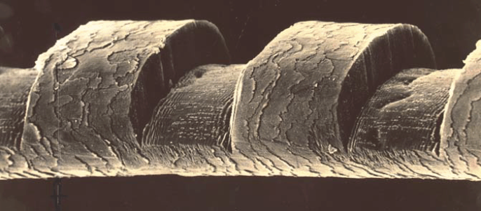 |
| Fig. 1 – Scanning electron micrograph of a human hair etched by irradiation with an ArF excimer laser; the notches are 50 ƒÝ wide. |
While IBM Intellectual Property Law was preparing a patent application, we were constrained from discussing our discovery with people outside IBM. But we had a newly hired IBM colleague, Ralph Linsker, with an M.D. and a Ph. D. in physics. Linsker obtained fresh arterial tissue from a cadaver, and we irradiated a segment of aorta with both 193-nm light from the ArF excimer laser and 532-nm light from the Q-switched, frequency-doubled Nd:YAG laser. Once again, the morphology of the tissue adjacent to the irradiated/incised regions, examined by standard tissue pathology techniques, was stunningly different, with irradiation by the 193-nm light showing no evidence of thermal damage to the underlying and adjacent tissue.
This experimental study on freshly excised human tissue confirmed that excimer laser surgery removed tissue by a fundamentally new process. Our vision–that excimer laser surgery would allow tissue to be incised so cleanly that subsequent healing would not produce scar tissue–was more than plausible, it was likely, subject to experimental verification on live animals.
First public disclosure
After IBM filed our patent application on December 9, 1982, we were authorized to publicly disclose our discovery. We wrote a paper and submitted it to Science magazine, but it was rejected, because one of the referees argued that the irradiation of living humans and animals with far ultraviolet radiation would be carcinogenic, making our laser surgical technique more harmful than beneficial. Since Srinivasan now had an invitation to give a presentation about his work on polymers at the upcoming CLEO 1983 conference in Baltimore, MD, co-sponsored by the Optical Society of America, we wanted to get a publication into print as soon as possible, so we resubmitted our paper to the trade journal Laser Focus, including some remarks about the new experiments on human aorta. Serendipitously, the Laser Focus issue containing our paper was published simultaneously with CLEO 1983, where, on May 20, Srinivasan gave an invited talk entitled “Ablative photodecomposition of organic polymer films by far-UV excimer laser radiation.” In this talk, he gave the first oral public disclosure that the excimer laser cleanly ablated biological specimens as well as organic polymers.
From excimer laser surgery to ArF excimer laser-based refractive surgery
At this very same CLEO 1983 meeting, on May 18, Stephen Trokel and Francis L’Esperance, two renowned ophthalmologists, gave invited talks on applications of infrared lasers to ophthalmic surgery. I attended both of their talks and was amazed at the results they obtained in successfully treating two very different ophthalmic conditions. I was well aware that the ruby laser was first used to eradicate a retinal lesion in late 1961, and retinal surgery with lasers had become widespread in the ensuing two decades, in particular to repair retinal tears and to treat diabetic retinopathy. But these treatments required a laser at a wavelength for which the ocular media anterior to the retina was transparent. Excimer laser light would be absorbed in a thin layer upon entering the cornea, so the excimer laser would be useless for treating retinal maladies.
But Trokel knew of ophthalmic conditions, such as the refractive imperfection known as myopia, that could be corrected by modifying the corneal curvature. A treatment known as radial keratotomy (RK), developed in the Soviet Union and being practiced in the United States, corrected myopia by using a cold steel scalpel to make radial incisions at the periphery of the cornea. When these incisions healed, the curvature of the front surface of the cornea was reduced, with the consequence that the patient’s myopia was also reduced. The technique could rarely give the patient uncorrected visual acuity of 20/20, but the patient’s myopia was definitely reduced. One serious drawback of RK was that the depth of the radial incisions left the cornea mechanically less robust, and the healed eye was more susceptible to “fracture” under impact, such as might occur during an automobile collision. Trokel speculated that the excimer laser might be a better scalpel for creating the RK incisions.
Upon learning of our discover of excimer laser surgery, Trokel, who was affiliated with Columbia University’s Harkness Eye Center in New York City, contacted Srinivasan and subsequently brought enucleated calf eyes (derived from slaughter) to our IBM Research Center on July 20, 1983. Srinivasan’s technical assistant, Bodil Braren, participated in an experiment using the ArF excimer laser to precisely etch the corneal epithelial layer and stroma of these calf eyes. The published report of this study is routinely referred to by the ophthalmic community as the seminal paper in laser refractive surgery.
To conduct studies on live animals, the experiments were moved to Columbia’s laboratories. Such experiments were necessary to convince the medical community that living cornea etched by the ArF excimer laser does not form scar tissue at the newly created surface and the etched volume is not filled in by new growth. The first experiment on a live rabbit in November, 1983, showed excellent results in that, after a week of observation, the cornea was not only free from any scar tissue, but the depression had not filled in. Further histological examination of the etched surface at high magnification showed an interface free from detectable damage.
L’Esperance, also affiliated with Columbia’s Harkness Eye Center, thought beyond RK and, in November, 1983, filed a patent application describing the use of excimer laser ablation to modify the curvature of the cornea by selectively removing tissue from the front surface, not the periphery, of the cornea. His U.S. Patent No 4,665,913 specifically describes this process, which was later named photorefractive keratectomy (PRK).
Soon ophthalmologist around the world, who knew of the remarkable healing properties of the cornea, were at work exploring different ways to use to excimer laser to reshape the cornea. From live animal experiments, they moved to enucleated human eyes, then to blind eyes of volunteers, where they could study the healing. Finally, in 1988, a sighted human was treated with PRK (Editors Note: by Dr. Marguerite McDonald at LSU), and after the cornea had healed by epithelialization, this patient’s myopia was corrected.
To read the complete article written by Dr. Wynne, including his footnotes, please follow this link.
What is Anisometropia?
Anisometropia can be a result of induced (created) myopia from a scleral buckle and/or cataract. If this is the case, it is usually correctable.
New Website for FOV and Webinar!
I have started another website dedicated to the treatment of floaters. Two webinars are also scheduled for January 2014. Sign up…they’re free
Annual AAO Meeting and Young Ophthalmologists
I just got back from teaching Young Ophthalmologists and other doctors about the need to communicate effectively, market and exhibit transparency through…a web page.
AMD Update 25: Results of The AREDS2 HOME Study of Notal Vision’s Home Monitoring Device for AMD Announced
In April of 2010, I wrote about the inclusion of Notal Vision’s ForseeHome AMD Monitor in the AREDS2 clinical trial.
The overall objective of the two arm randomized clinical trial was to determine if home monitoring of participants at high risk of progression from late-stage dry AMD to neovascular AMD, using the comprehensive visual field and telemedicine solution based on the ForeseeHome Device in AREDS2 (referred to as the ForeseeHome comprehensive solution), would improve detection of progression to choroidal neovascularization (CNV) when compared with standard care (may have included use of the Amsler Grid).
Well, the results are in and the National Institute of Health (NIH) found that patients at high-risk for developing neovascular age-related macular degeneration would benefit from using the ForeseeHome Monitoring device for early detection of their CNV.
Report Represents the Most Comprehensive Study of Home Monitoring for Progression of AMD
As reported at the Retinal Subspecialty Meeting at this year’s AAO Meeting, the results of the Home Monitoring of the Eye (HOME) study, conducted in Age-Related Eye Disease Study 2 (AREDS2) clinical centers showed that participants at high risk for developing choroidal neovascularization (CNV) using the ForeseeHome monitoring device strategy had significantly better preservation of their visual acuity at the time of CNV detection than the control group of participants who were only using standard care methods (the Amsler grid) to self monitor their AMD for progression. The study’s Data Safety and Monitoring Committee recommended early termination of the study on April 2, 2013 based on superior vision outcomes among the participants randomly assigned to use the home device.
The AREDS2 HOME Study was a collaborative effort led by the National Eye Institute to evaluate the performance of a home monitoring device plus standard care compared to standard care monitoring alone for the detection of AMD progression to the neovascular phase. Standard care methods included periodic monocular self checks of vision clarity, blind spots and distortion, which included use of an Amsler grid. As treatments to manage the neovascular phase of AMD have improved, the importance of early detection of this event has increased in an effort to optimize outcomes following treatment of neovascular AMD. Approximately 8 million individuals in the United States, age 50 and older, are estimated to have intermediate (large drusen) or advanced dry AMD in one eye, placing them at high risk of progression to neovascular (wet) AMD (CNV), ranging from 25 to 50% over a five-year period.
Results of the HOME Study and Implications for AMD Management
At the time of CNV detection, 87% of eyes in the ForeseeHome device arm maintained visual acuity of 20/40 or better compared to 62% in the standard care alone arm. Median acuity among device users at the time of CNV diagnosis was 20/32. Among participants who used the device at the recommended minimum frequency (twice per week) to monitor their AMD for progression, 94% of eyes that progressed to CNV maintained 20/40 or better visual acuity. When CNV was detected, participants in the ForeseeHome device arm lost fewer letters on visual acuity testing (median loss of 4 letters) from entry levels of vision at the start of the study compared to those in the standard care alone arm (median loss of 9 letters). Use of the ForeseeHome device resulted in an increase in the proportion of CNV events first identified at home, meaning in between routine ophthalmic office visits to assess detection of disease progression. Among individuals using standard care methods for monitoring, only 55% of those that progressed noted symptoms at home that led them to present for examination; whereas 80% of the participants in the device monitoring group returned sooner than a scheduled visit because a change was noted by the device or by self-monitoring. This was associated with a greater degree of vision preservation at CNV diagnosis among individuals who returned promptly for changes, as the median visual acuity loss at CNV detection was 3.0 letters for those in the device arm compared with 11.5 letters for those in the standard care group. The average annual rate of false alerts among the device users, reported as the annual false positive rate, was 0.24 alerts/year, which may be extrapolated to one false alert on average every 4.2 years for each ForeseeHome user.
“Persons 60 years of age or older should undergo dilated eye examinations to determine their risk of developing advanced AMD, especially CNV,” said Jeffrey S. Heier, MD, Director of the Vitreoretinal Service and the Director of Retina Research at Ophthalmic Consultants of Boston and one of the principal investigators of the HOME Study. “In contrast to current home monitoring strategies, those with intermediate AMD (bilateral large drusen) or advanced AMD in 1 eye are likely to benefit from home monitoring with the ForeseeHome device to detect the development of CNV at an earlier stage with better preservation of their visual acuity to maximize visual acuity results after intravitreal therapy with anti-VEGF agents.”
About the HOME Study
The HOME Study was a controlled, randomized clinical trial that was part of AREDS2. The study was conducted in 44 clinical centers across the U.S., enrolling 1,520 participants at high risk for developing CNV. (With approximately half using the device and the other half acting as controls, using standard care.) The objective of the HOME Study was to determine whether monitoring with the ForeseeHome device plus standard care results in earlier detection of CNV compared to standard care alone. Standard care included instructions to the patient on self-monitoring for CNV. Better visual acuity at the time of CNV detection is both a reflection of earlier CNV detection as well as a favorable predictor for visual function outcomes following the management of CNV with intraocular anti-angiogenic medications.
About the ForeseeHome AMD Monitoring Program
The ForeseeHome AMD Monitoring Program is a prescription-based, comprehensive telemonitoring and data management system that extends the management of AMD to patients’ homes between office visits. The test results are transmitted to a central monitoring center that will alert, physicians to immediate, significant visual field changes in their patients, so that patients can be recalled for timely follow-up and necessary treatment may be initiated. The ForeseeHome AMD Monitoring Program utilizes a simple to use device based on preferential hyperacuity perimetry, a form of visual-field testing, to identify minute visual distortions, or metamorphopsia, for the detection of early CNV development.
To read more about Notal Vision and the ForseeHome device, read my full report of March 9, 2010: Notal Vision: The ForeseeHome AMD Monitor and It’s Potential to Save Vision – A First Report.
AMD Update 24: DARPins Phase 2 Trial Results Fall Short
Back in February, I first reported on Allergan’s DARPins in my Update 23: DARPins, The Next “Game Changer” for Wet AMD? In that report, I wrote that Molecular Partners’ MPO112 (Allergan’s AGN-150998) showed promise of improving vision and having a long ocular half-life which appeared to be a vast improvement over both Lucentis and Eylea, perhaps requiring injections every 3-4 months compared to bi-monthly for Eylea and monthly for Lucentis and Avastin. (I also noted a second agreement with Molecular Partners, the licensors of the DARPin technology to Allergan, in which a combination dual action anti-VEGF/PDGF drug therapy was also be under investigation.)
Well, the first part of the promise, the longer interval injection rate for the DARPins, has fallen through. As reported by two analyst groups, Allergan presented results last Friday (November 15th) from the Phase 2 trial of AGN-150998 (anti-VEGF DARPin program) in wet AMD at the Retina Subspecialty Meeting ahead of the start AAO annual conference in New Orleans. The results supported the company’s decision several months ago, to slow down advancement of the clinical trial, in that the drug failed to meaningful delay the time to retreatment and the associated rates of inflammation were higher than were anticipated. Though Allergan continues to evaluate the drug and still may ultimately advance it into Phase 3 studies, there appears to be only limited competitive threat to Eylea (or, perhaps Fovista, Ophthotech’s combination anti-VEGF/PDGF drug in clinical study – see my two write ups on Fovista, shown below, for more information about this potential drug). Specifically, the analysts see a low likelihood of commercial adoption or integration into the treatment paradigm for wet AMD without any sustained improvement in visual acuity or meaningful delay in the time to retreatment.
In looking at the data presented, the study evaluated two doses of the AGN-150998 (3mg and 4.2mg) vs. Lucentis. The drug was administered at week 4 and then pro re nata (PRN) or by week 16, and then again PRN or by week 32 at the latest. At day 60 and day 90, the 4.2mg dose appeared to delay the need for retreatment in ~10-15% of patients. Looking at the data another way, the 4.2mg dose appeared to delay the median time to retreatment by ~20 days. There were no differences in the percent of patients gaining 15 or more letters in best corrected visual acuity (BCVA) from baseline by week 16, and again at week 32.
In terms of safety, the AGN-150998 treatment was associated with a meaningful rate of ocular inflammation adverse events relative to Lucentis (13% vs. 0%). Specifically, treatment with AGN-150998 had higher rates of uveitis (3% with 3mg, 6% with 4.2mg, 0% with Lucentis), anterior chamber inflammation (2% and 3% vs. 0%), vitritis (7% and 2% vs. 0%). For reference purposes, historical data imply the rate of intraocular inflammation in AMD trial are 13% and 1% with Lucentis and Eylea, respectively.
Allergan has indicated that it would be making changes to the manufacturing process to hopefully reduce the inflammation seen in the Phase 2 trials, when and if they decide to proceed to a Phase 3 trial.
I was not able to determine if Allergan and Molecular Partners still plan to go ahead with a clinical trial for the dual action drug, which remains in a pre-clinical stage.
References:
Analyst Reports – Private correspondence.
Fovista Reports:
Giant Retinal Tears
Giant retinal tears usually lead to blindness. I use a combined operation with Perfluoron where this tends not to happen…and there’s a video!
Preventing Giant Retinal Tears
Here’s a video and post about preventing the formation of a Giant Retinal Tear….in a patients other eye!
A Branch Retinal Vein Occlusion: aka BRVO
A branch retinal vein occlusion reduces vision by causing macular edema. Treating the macular edema may improve vision.
Gene Therapy in Ophthalmology Update 21: New Gene Therapy Company, Spark Therapeutics, Launches
Children’s Hospital of Philadelphia (CHOP) announced that it had spun off its work in gene therapy to a new, fully integrated company, Spark Therapeutics, that will assume control over two current gene therapy clinical trials: a Phase III study for Leber’s Congenital Amaurosis, an inherited disease that results in blindness caused by mutations of the RPE65 gene, and a Phase I/II study for hemophilia B. The new company is also advancing toward the clinic with gene therapy programs to address neurodegenerative diseases and additional hematologic disorders and other forms of inherited blindness. One such program, in the latter category, already in pre-clinical development at CHOP, could be its study for the treatment of Choroideremia, a rare inherited disorder that causes progressive loss of vision due to degeneration of the choroid and retina.
Editors Note: It should be noted that one clinical trial using gene therapy to treat Choroideremia is already underway at Imperial College London and Oxford University, in conjunction with Moorfields Hospital in London.
The new company has been launched with a $50 million capital commitment from Children’s Hospital to advance and commercialize multiple ongoing programs with clinical proof of concept.
As noted by Susan Young, writing about the launch in Technology Review, “Spark has a chance to be the first gene-therapy company to obtain FDA approval. Results for a late-stage trial of a gene therapy for Leber’s Congenital Amaurosis … are expected by mid-2015. That treatment is one of several gene therapies in or nearing late-stage testing contending to be the first gene therapy approved by the FDA for sale in the U.S.”
The Phase III trial was initiated late last year, and CHOP has made significant progress in enrolling patients. Spark will be sharing additional details on its progress and encouraging results in the very near future.
And, as shown in my table of Ongoing Clinical Trials in Ophthalmology, it is the only gene therapy trial (in ophthalmology) that has advanced to Phase III.
Editors Note: For clarity, is should be noted that there are five other clinical trials underway to treat Leber’s, as shown in my table, but all are currently Phase I or Phase I/II studies. The Spark Therapeutics trial is the farthest advanced.
“The creation of Spark is the culmination of a decade-long commitment by CHOP and our founding team to drive the field of gene therapy forward during a time when many in the industry had moved away,” said Jeffrey D. Marrazzo, co-founder, president and chief executive officer of Spark Therapeutics. “Their vision and long-term dedication have enabled us to effectively address many of the key challenges facing the field and to emerge with one of the industry’s most robust clinical-stage gene therapy pipelines; as well as exclusive rights to commercialize a proprietary manufacturing platform, supply from a world-class manufacturing facility and a founding team with a proven track record of executing safe and effective gene therapy trials for nearly two decades. We are working with great urgency and care to deliver gene therapy products with the potential to transform the lives of those affected by severe genetic diseases.”
Spark builds on the work of CHOP’s Center for Cellular and Molecular Therapeutics (CCMT), established in 2004 as a world-class center for gene therapy translational research and manufacturing. Many of the CCMT’s leaders will assume management roles within Spark or engage with the company as scientific advisors, including Katherine A. High, M.D, a gene therapy pioneer who has served as the director of the CCMT since its inception.
“Gene-based medicines are among the most complex therapeutics ever developed,” said Dr. High. “We at CCMT have persevered through more than a decade of scientific and clinical development and are now closer than ever to realizing the ambitious vision of one-time, potentially curative therapies to address serious genetic conditions. The team at Spark has incredible goals for the treatment of diseases including hemophilia B and inherited blindness, and we look forward to working with them to deliver groundbreaking new treatments to patients in need.”
Spark has entered into agreements with multiple academic institutions to assemble the technology, programs and capabilities needed to deliver its pioneering gene therapy products. Notably, Spark has exclusive rights to commercialize CHOP’s proprietary manufacturing technology and will use clinical-grade gene therapy vectors produced by the CCMT’s state of the art good manufacturing practices (cGMP) clinical facility.
Pioneers in AAV delivery
Over the past two decades, the Spark leadership team has developed unrivaled expertise in the design, manufacturing and delivery of gene therapies using adeno-associated virus (AAV) vectors. AAV has been demonstrated in clinical studies to be a safe and effective vehicle for the delivery of genetic material into targeted cells and provides unique advantages over alternative delivery approaches. The Spark team was among the first to demonstrate human clinical proof of concept in two distinct organ systems — the eye and the liver — establishing a strong foundation for the company’s current programs, and has clinical experience in 15 studies across diverse genetic and non-genetic diseases and five distinct routes of administration.
Spark’s most advanced clinical program is a Phase III study to address blindness caused by mutations in the RPE65 gene. There is currently no pharmacologic treatment for this form of inherited retinal degeneration, which ultimately causes irreversible blindness.
The open-label, randomized, controlled study builds on an earlier clinical study in which 12 patients with RPE65-related blindness demonstrated notable improvement in visual function, moving in some cases from being profoundly blind to being able to recognize faces and ambulate independently. All school-age patients enrolled in the trial were able to transfer from Braille classrooms to sighted classrooms.
One such patient was Corey Haas, whose story is related in the book “The Forever Fix: Gene Therapy and the Boy Who Saved It”.
 |
|
| Corey Haas, his parents, and the CHOP team that treated his Leber’s and gave him back his vision. |
 |
| Read Corey’s story in Ricki Lewis’ book, The Forever Fix: Gene Therapy and the Boy Who Saved It. |
The team’s experience in the clinical study of gene therapy – from designing and manufacturing vectors to conducting studies that have shown strong potential for safety and efficacy – is unparalleled in the field. Clinical-grade vectors prepared by the team have been used to safely treat more than 100 human subjects in 12 clinical trials in the U.S. and EU, across five parenteral routes of administration in genetic and non-genetic diseases. No other group can claim this breadth of expertise and experience in human gene therapy.
The adeno-associated virus (AAV) vectors used in the clinical programs have been demonstrated to be safe and effective vehicles for delivering genetic material into targeted cells, providing unique advantages over alternative therapeutic approaches. The team has established human proof of concept in two organ systems – the eye and the liver – and are advancing a Phase III program in blindness caused by mutations of the RPE65 gene; a Phase I/II program in hemophilia B; and preclinical programs in neurodegenerative diseases and other hematologic disorders and forms of inherited blindness.
What is a Choroidal Nevus
You’ve been diagnosed with a “freckle.” A choroidal nevus is benign and common,but malignant transformation can occur.
Gene Therapy in Ophthalmology Update 20: Oxford BioMedica Clinical Trials Resume
Back in June, Oxford BioMedica announced that it had voluntarily paused recruitment for its clinical trials for wet AMD (RetinoStat Phase I), Stargardt’s Disease (StarGen Phase I/IIa) and Usher’s Syndrome (UshStat Phase I/IIa). The company had halted recruitment of the aforementioned studies, as a precautionary measure, while it investigated the detection of very low concentrations of a potential impurity in its clinical trial material derived from a third party raw material.
Oxford has since performed extensive characterization studies using its newly developed, state-of-the-art analytical methods to identify the impurity as highly fragmented DNA derived from fetal bovine serum (FBS), the most widely-used growth supplement for cell culture media. In light of these findings, Oxford remains convinced of the safety, integrity and quality of its LentiVector platform products and no safety concerns relating to any of the ocular products have been identified in any pre-clinical and clinical data generated to date.
Today, the company announced that following the submission of a comprehensive data package to the FDA and the French regulatory agency, ANSM, it has received agreement from both agencies to resume recruitment into its ocular clinical trials using the existing clinical trial material. The company will continue to use highly sensitive, state-of-the-art analytical methods to ensure the quality and integrity of its lentiviral vector products and will work with FDA and ANSM to define the necessary specifications for future batches of clinical trial material.
Oxford is now working closely with the clinical trial centers to obtain the necessary ethics committee approvals in order to resume recruitment into the clinical studies.
(For a list of the clinical site centers in the U.S. and France involved in the three studies, please take a look at my Gene Therapy Ongoing Clinical Trial Table at http://tinyurl.com/GeneTherapyClncal)
John Dawson, Chief Executive Officer of Oxford BioMedica, said: “We value our relationships with the regulatory authorities and are pleased that, on the basis of our extensive technical investigations to demonstrate the integrity of our products, FDA and ANSM agree with our proposal to resume treating patients in our ocular trials as soon as possible.
“We place the highest importance on safety, and our analytical methods and quality assurance processes are continuously evolving to ensure that we remain at the forefront of gene therapy development and manufacture. I am confident that, with significant opportunities ahead such as the recently-announced AMSCI project win, Oxford BioMedica will continue to lead the way in delivering novel gene therapies to patients.”
For your information, Oxford BioMedica has reported that 9 of the 18 patients to be treated in the wet AMD clinical trial had been treated; 12 of the 28 patients in the Stargardt’s trial; and 3 of 18 patients in the Usher Syndrome trial had been treated prior to the halt in recruitment in June.
Coincidently, Genzyme, who is also running a gene therapy clinical trial to treat the wet form of AMD, also announced a halt in recruitment for its trial in July. No reason for the stoppage has been given and all attempts to determine why the halt in recruitment occurred have been rebuffed. As of the last time I had obtained reliable information about the Genzyme trial, 6 of 34 patients to be treated had been treated.
When to Remove Macular Pucker
Most patients wait too long before considering removal of a macular pucker (epiretinal membrane). The decision should be made as soon as visual changes are noticed.
What is the Vitreous?
The vitreous causes most surgical diseases of the retina. If everyone had a vitrectomy (to remove the vitreous), many people retinal patients couldn’t lose vision!
Happy Birthday to Me….
Here are few random thoughts on turning 53 and the events of the past year. Hope you are well.
Sophie is 93 and Reads!
I’ve written about another of my more elderly patients who remains independent and can read despite wet ARMD! She’s 93!
Research in Retinal Disease: The Foundation Fighting Blindness Invests $2.1 Million in Seven New Research Efforts
As I continually search the web for interesting news about new technologies for treating retinal diseases, I came across this news from the Foundation Fighting Blindness’ website yesterday afternoon. It relates to some of the annual grants to researchers that the FFB will be funding this year. It includes better ways of looking at retinal cells (via use of the adaptive optics laser scanning ophthalmoscope) and several projects involving gene therapy, along with a couple looking at ways of, hopefully, stopping the progression of dry AMD.
The following write up is reprinted with permission of the FFB.
August 15, 2013
The Foundation’s Scientific Advisory Board (SAB) recently completed its annual grants review process, leading to the allocation of $2.1 million in funding for seven new research projects, including those for identifying new disease-causing gene mutations, developing cross-cutting gene therapies and advancing potential treatments for dry age-related macular degeneration. The three-year grants were awarded after the SAB reviewed 117 proposals submitted to the Foundation last October.
“Grants review is a rigorous, multi-step process that takes most of the year to complete,” says Stephen Rose, Ph.D., chief research officer, Foundation Fighting Blindness. “Due to revenue limitations, we can only fund a fraction of the high-quality projects we’d like to fund. That makes the selection process even more challenging. We had to leave several excellent proposals on the table.”
Here are brief descriptions of the new research projects:
AOSLO: Detecting Retinal Degeneration Before Vision is Lost
The adaptive optics laser scanning ophthalmoscope (AOSLO) is like a powerful microscope that enables retinal researchers to see structural changes in the retina well before vision is lost from a retinal disease. That power can enable researchers to more quickly determine if a treatment is working in a clinical trial. Austin Roorda, Ph.D., of the University of California, Berkeley, is performing studies of AOSLO to correlate changes in the retina (e.g., loss of photoreceptors) with changes in vision.
Enhancing AOSLO for Expanded Clinical Use
Like Dr. Roorda, Stephen Burns, Ph.D., of the University of Indiana, is working with AOSLO to study the correlation between retinal and vision changes. He is also making AOSLO more affordable by using newer camera technology. In addition, he’s employing state-of-the-art computing technologies derived from video games to decrease image-processing times and costs. The new technology will make the imaging process more comfortable for the patient by tolerating more head and eye movement.
Figuring Out Why Severity of Vision Loss Varies for People with XLRP
Researchers have reported for many years that the severity of vision loss for people with X-linked retinitis pigmentosa (XLRP) can vary greatly, even for people within the same family. Stephen Daiger, Ph.D., of the University of Texas Health Science Center at Houston, will be looking at the role of a various biological, genetic and environmental factors in vision-loss variability for those with XLRP. The identification of a significant factor that modulates vision-loss severity – perhaps a protective protein – could lead to a potential treatment.
Finding New Genes Linked to ADRP
Researchers have identified almost two dozen genes linked to autosomal dominant retinitis pigmentosa (adRP), but many are yet to be found. Rui Chen, Ph.D., of Baylor College of Medicine, is on the hunt for those remaining adRP genes. With DNA from 118 adRP families, including 18 families with at least nine affected members, Dr. Chen is well positioned to identify additional genes linked to adRP. Finding the new genes will provide researchers with targets for treatments and cures.
Developing Neuroprotective Gene Therapies to Preserve Vision
John Ash, Ph.D., is developing gene therapies that have the potential to preserve vision in people affected by a broad range of retinal diseases. Unlike corrective gene therapies, which work only for conditions caused by a specific gene, Dr. Ash’s proposed treatments are designed to keep the retina healthy independent of the underlying disease-causing gene. He also believes the proteins delivered by his treatments – PIM-1 and STAT3 – will be less likely to cause damaging inflammatory side effects than some previously investigated neuroprotective proteins.
Targeting Inflammation to Halt AMD
Thanks to previous Foundation-funded genetic studies, researchers have strong evidence that the progression of age-related macular degeneration is associated with an over-active immune system. This ultimately leads to inflammation and cell death in the retinal pigment epithelium (RPE), a layer of cells that provides critical waste and nutritional support to photoreceptors. Loss of the RPE subsequently leads to loss of photoreceptors and vision. Jayakrishna Ambati, M.D., of the University of Kentucky, is developing a gene therapy that preserves the RPE by preventing the harmful sequence of immune-system events.
Boosting Cells’ Energy Supplies to Save Vision in AMD
Based on prior research, Deborah Ferrington, M.D., of the University of Minnesota, believes that mitochondrial dysfunction in the RPE plays a significant role in the development of AMD. Mitochondria are like miniature organs (organelles) within all cells that provide energy. When not working properly in retinal cells, they can lead to cell death and vision loss. Dr. Ferrington is evaluating compounds that help protect mitochondrial function in the RPE.