Category: Blogs

A New Approach to Treating Chronic Kidney Disease: Replenishing Your Nephrons

As some of you may know, I have chronic kidney disease (CKD). Thanks to my wife and the scare put into me by my nephrologist, that I would soon have to begin dialysis, I have managed to get my CKD  in remission, or at least under control. My GFR number (that indicates when you must start dialysis) has held steady, or actually gotten better since my wife put me on a strict diet and I have lost about 15 pounds – and my nephrologist took me off of lisinopril (for blood pressure control), which seemed to raise my GFR by about three points. So, it appears I won’t have to go on dialysis any time soon.
All that being said, and with my strong interest in the use of stem cells and gene therapy in treating retinal diseases in ophthalmology, I have been searching the web for research on the use of stem cells to treat kidney disease or to produce new nephrons, the kidney cells that filter the blood as it passes through the kidney – and that go bad or die causing CKD. I think I have found very early research of that possibility.
Earlier this week, I saw a news  release from the University of Queensland in Australia that said that Dr. Melissa Little and her research group at the Institute of Molecular Bioscience (IMB) have found a set of six genes that can prompt some types of adult kidney cells to regress to an earlier stage of development (stem cells) and act like the precursors to the cells of the nephron. Since it is death or damage of nephrons that causes chronic kidney disease, by forcing adult cells to act like early nephrons, they may have potentially found a way to trigger the growth of new filters in the kidney.
All of your nephron cells are formed before birth and people with fewer nephrons are at higher risk of kidney disease.
Note: Dr. Little is one of sources that I had found in my web search as she had published a paper on “Stem Cell Options for Kidney Disease” in 2008, and we had corresponded at the beginning of this year when I began my search for a stem cell answer.
“This discovery is the first of its kind and offers hope to patients with chronic kidney disease. If we can find a way to provide new nephrons to an adult or increase nephron numbers in babies at birth, we could potentially reduce the risk of disease progression,” said Professor Little.
This landmark paper, “Direct Transcriptional Reprogramming of Adult Cells to Embryonic Nephron Progenitors”, by Caroline E. Hendry, Jessica M. Vanslambrouck, Jessica Ineson, Norseha Suhaimi, Minoru Takasato, under the supervision of Professors Fiona Rae and Melissa H. Little, was published June 14th in the Journal of the American Society of Nephrology, the world’s leading nephrology journal.
Professor Little said, “There was still more work to be done to encourage these reprogrammed early nephron cells to function and integrate. While this is a beginning, we hope it will inspire industry leaders and researchers around the world to invest further in cellular and bioengineering approaches to kidney repair and regeneration.”
Stem Cells Australia Program Leader and Chair of Stem Cell Science at The University of Melbourne, Professor Martin Pera welcomed the research findings. “This innovative study provides evidence that adult cells can be reprogrammed to resemble the cells in the embryo that give rise to the kidney. The results pave the way for future studies that will enable researchers to produce human kidney cells in the laboratory, for use in studies of renal disorders, and for testing new drugs. Eventually this technology might help to make cells for transplantation to treat kidney disease,” said Professor Pera.
The Technology
I have attempted to read Dr. Little’s paper on reprogramming kidney cells, and with her assistance, this is what I understand she and her colleagues have done, which is a very early step in the long road to someday being able to replenish nephron cells in an adult kidney.
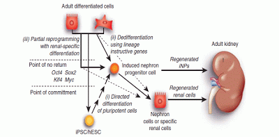
In an earlier paper written by Caroline Hendry and Dr. Little, “Reprogramming the kidney: a novel approach for regeneration”, they discussed the various approaches that might be taken to re-create viable cells within a diseased kidney, including using  induced pluripotent stem cells (iPSCs) derived from skin cells or other sites, or even the use of embryonic stem cells (ESCs) that would be introduced into the kidney to form new nephrons (?), if they could – as shown in the accompanying figure (but how would you control the formation of the new cells?). But they concluded that the best approach would be reprogramming existing kidney cells to the progenitor stage, with the hope that these would develop into the needed new cells, or in this case, nephrons, the approach they ultimately used in this new research.
Figure1 | The application of reprogramming to the kidney, indicating the feasible starting cells and target phenotypes.
(i) Reprogramming may involve the directed differentiation of human embryonic stem cells (hESCs)/induced pluripotent stem cells (iPSCs) to a renal lineage. The iPSCs may be recipient-derived and may be derived from adult kidney cells or any other available adult cell type using the same factors. Directed differentiation is likely to recapitulate development; hence, it is likely to require differentiation through a nephron progenitor intermediate (induced nephron progenitor cell; iNP) but may continue on to more specific mature renal cell types.
(ii) A specific renal lineage may also be achieved via lineage-instructive reprogramming directly to that state from an adult cell type. Again, this may be the renal epithelium, renal stroma, or any other available differentiated adult cell type; however, this is likely to be more successful if the attractor states of the starting and target cell type are as close as possible. Reprogramming may be to the iNP state or directly to a more mature renal cell fate.
(iii) Finally, reprogramming may use the classical Yamanaka factors until the cells pass the point of no return, after which a renal lineage may be reached via the application of the appropriate environmental cues. Such cues may once again target the iNP state or aim to directly induce a more mature renal cell type.
As previously stated, the nephron progenitor population of the embryonic kidney gives rise to all of the nephron cells that will be present in the adult kidney, prior to birth. So, currently, what you’ve got at birth is what you live with.
Using a screening technique, the researchers were able to identify a group of six genes, that activate a network of genes that can reprogram adult proximal tubule cells back to the nephron progenitor stage – which in turn can form adult nephron cells. Although the researchers believe that other factors are required, they concluded that these results suggest that re-initiation of kidney development (nephron cells) from a population of adult cells (proximal tubule cells) by generating embryonic progenitors may be feasible, opening the way for additional cellular and bioengineering approaches to renal repair and regeneration.
In their literature search, they could not find any previous reports of kidney cells being reprogrammed back to a progenitor cell type. Their hope is that this discovery will lead others to follow their lead and begin further work in the possible reprogramming of adult kidney cells for the repair and rejuvenation of diseased kidneys.
Sources:
News Releases:
Research reprograms future of kidney health, Institute for Molecular Bioscience and Stem Cells Australia, June 14, 2013
Papers:
Reprogramming the kidney: a novel approach for regeneration, Hendry and Little, Kidney International, March 21, 2012
Stem Cell Options for Kidney Disease, Hopkins et al, Jnl of Pathology, October 20, 2008

Gene Therapy in Ophthalmology Update 19: A New Virus Vector for Safer Delivery of Gene Therapies

Researchers at the University of California at Berkeley, along with some assistance from the Flaum Eye Institute and Center for Visual Science at the University of Rochester, have come up with a new version of an adeno-associated virus (AAV) vector that can deliver genes deep into the retina using an intravitreal injection of the vector into the vitreous, a less-invasive technique, instead of an intraretinal injection below the surface of the retina, which is the common way gene therapy is currently delivered.
The study was authored by postdoctoral fellows Deniz Dalkaral (then with Helen Wills Neuroscience Institute of UCal Berkeley, but now with Institut de la Vision in Paris) and Leah C. Byrne (Helen Wills), and graduate students Ryan R. Klimczak and Meike Visel (UCal Berkeley’s Dept. of Molecular and Cell Biology), and Lu Yin and William H. Merigan (Flaum Eye Institute and Center for Visual Science at the Univ. of Rochester), under the direction of Professors John G. Flannery, and David V. Schaffer of UCal Berkeley. The paper, “In Vivo–Directed Evolution of a New Adeno-Associated Virus for Therapeutic Outer Retinal Gene Delivery from the Vitreous”, was published online on June 12th in Science Tranlational Medicine.
As explained by Dr. Jean Bennett, a professor of ophthalmology at the University of Pennsylvania in Philadelphia, who was not involved in the study, but who has done extensive work with gene therapy in the treatment of Leber’s congenital amaurosis, “It shows the results of a very clever system to evolve AAV to target cells in the retina efficiently from an intravitreal injection.”
“Intravitreal injection, whereby a needle is pushed into the eye’s vitreous, or gel-like core, is a common drug delivery procedure performed under local anesthetic in a doctor’s office”, explained Bennett. “But using this routine injection technique in trials of gene therapy for retinal degeneration has thus far proven impossible.”
The problem, as explained by Dr. David Schaffer, a professor of chemical and biomolecular engineering, bioengineering, and neuroscience at the University of California, Berkeley, who led the research, is that current AAV vectors are incapable of penetrating deep into the retina where the target cells for retinal diseases are located. “AAV is a respiratory virus and so it evolved to infect lung epithelial cells,” explained Schaffer, “It never evolved to penetrate deep into tissue.”
Patients receiving gene therapy have theretofore undergone a vitrectomy (removal of the vitreous) and a direct intraretinal injection, which requires hospitalization and general anesthetic, and can sometimes even damage the retina. “If it were possible to inject AAV into the vitreous instead of the retina and still get gene delivery to the target cells, said Bennett, “one could envision the [doctor saying], ‘Ok, well just come into the office and get your gene therapy, tomorrow afternoon at two.’”
With that aim, Schaffer and colleagues used a process called “directed evolution” to randomly create millions of variations of the AAV virus to determine which ones were better at tissue penetration. They injected regular AAV into the vitreous of mouse eyes and one week later collected photoreceptor cells from deep within the retina. The tiny percentage of AAV vectors that made it into those cells were then amplified, repackaged into virus particles and injected into the vitreous again. They repeated the injection, recovery, and amplification a total of six times, finally isolating 48 AAV variants for sequencing. Two thirds of those isolates turned out to be the same variant, and Schaffer and colleagues named it 7m8.
Lastly, to determine whether the 7m8 vector would be likely to show similar deep penetration in the human retina, Schaffer had the vector fused to a fluorescent protein injected into the vitreous of macaque eyes. Primate retinas are considerably thicker than those of mice, and the vector did not consistently reach the deep cell layers – showing a spotty penetration pattern rather than the wide and even pan-retinal penetration that had been seen in the mice. However, 7m8 did effectively target photoreceptor cells of the fovea – a thinner part of the primate retina that is essential for the sharp detailed vision humans use when reading and driving. “That’s a really important region to protect,” said Schaffer. “For the quality of life of patients who are going blind, if you can at least protect the fovea that would be a huge improvement.”
Eye cells labeled with green fluorescent protein have successfully taken up the virus, showing that the ‘evolved’ virus (right) is more effective than the virus currently used for gene therapy (left). The new virus is particularly good at targeting the critical photoreceptors (top layer). (Source: University of California, Berkeley)
Note: All of the “directed evolution” work with mice to discover the 7m8 AAV vector was done at UCal Berkeley, while the confirmatory primate injection and imaging was done at the Univ. of Rochester.
Schaffer and colleagues don’t yet know what makes the 7m8 vector so much better at tissue penetration than its AAV ancestor, but they plan to find out and use that knowledge to further improve its penetration in the primate retina.
“Building upon 14 years of research, we have now created a virus that you just inject into the liquid vitreous humor inside the eye, and it delivers genes to a very difficult-to-reach population of delicate cells in a way that is surgically non-invasive and safe. “It’s a 15-minute procedure, and you can likely go home that day.”
The engineered virus works far better than current therapies in rodent models of two human degenerative eye diseases (X-linked retinoschisis and Leber’s), and can penetrate photoreceptor cells in monkeys’ eyes, which are similar to those of humans.
Schaffer said he and his team are now collaborating with physicians to identify the patients most likely to benefit from this gene delivery technique and, after some preclinical development, hope soon to head into clinical trials.
Schaffer predicts that the viruses can be used not only to insert genes that restore function to non-working genes, but can knock out genes or halt processes that are actively killing retinal cells, which may be the case in age-related macular degeneration.
As noted by  Dr. Stephen Rose, Ph.D., chief research officer, Foundation Fighting Blindness, one of the co-funders of the research, “This is a critical next step in the development of retinal gene therapies. The enhanced AAV holds potential for treating more of the retina and doing so more safely. Incremental advancements like this are essential to getting the best treatments out to the patients.”
The investigators showed efficacy for the 7m8 AAV in a large animal as well as mouse models of retinoschisis and Leber congenital amaurosis, or LCA (RPE65 mutations). In the mouse studies, the virus was able to penetrate the retina and deliver a corrective gene to enable the retina to function normally.
While the large animal did not have a retinal disease, the virus transduced many regions of its retina. Ultimately, in both types of animals, the AAV was able to deliver genetic cargo to a variety of retinal cells, including: photoreceptors, the cells that provide vision; the retinal pigment epithelium, a layer of cells providing nutrients and waste disposal; and ganglion cells, which are a target for emerging, vision-restoring optogenetic therapies. (Editor’s Note: See, for example, my writeup of the “Nirenberg Technique”, an optogenetic approach to restore near normal vision to the blind.)
Most notably, the intravitreally administered AAV was able to penetrate the fovea, a small pit in the center of the retina rich in cones, which provides the vision most critical to daily living, but is often made fragile by degenerative diseases. Researchers have been concerned that injections underneath the fovea could cause permanent damage and vision loss in patients with advanced degeneration in their central retina.
AAVs are currently used for gene delivery in several retinal gene therapy clinical trials, including those that have restored vision in children and young adults with LCA (RPE65). AAVs are attractive for gene delivery because of their natural ability to penetrate a variety of cells. In addition, humans are exposed to the virus in nature and, therefore, tolerate it well.
To identify the optimal AAV for intravitreal gene delivery, the scientists used a process called “directed evolution” to randomly create millions of variations of the virus. The variants were then screened in mice to identify the top candidates for gene delivery to the retina. In addition to looking for an AAV that could penetrate retinal cells well, the researchers searched for a variant that could pass through a formidable barrier in the eye known as the inner limiting membrane, or ILM, which separates the vitreous from the retina.
The scientists from UC Berkeley plan to perform additional toxicology and efficacy studies to ready the 7m8 AAV for study in humans.
Sources:

Researchers Identify Better Virus for Retinal Gene Delivery, Foundation Fighting Blindness, June 12, 2013

Stem Cells in Ophthalmology Update 25: ACT Patient in Dry AMD Trial Goes from 20/400 to 20/40!

The story started innocently enough. On Wednesday, May 15th, the journal Cell reported on a study that claimed biologists had finally created human stem cells by the same technique that produced Dolly the cloned sheep in 1996. They transplanted genetic material from an adult cell into an egg whose own DNA had been removed.
OK, an important story but what followed boggles the mind. Many science reporters wrote about the discovery which got picked up by several news sources. However, a sharp-eyed member of the Investor Stemcell Forum (iCell), a group originally started by investors in Advanced Cell Technology, noticed a quote at the end of Sharon Begley’s writeup. Sharon is a science reporter writing for Reuters. She had obtained quotes from several people working in the stem cell field, including at the very end of her story, the following quote from Dr. Robert Lanza, the Chief Scientific Officer of Advanced Cell Technology (ACT), “The most promising human study is ACT’s. It is two years into clinical trials using stem cells derived from human embryos to treat two forms of blindness, including macular degeneration, with encouraging results. One patient’s vision went from 20/400 to 20/40, said Lanza.
By Wednesday afternoon and into the evening, the iCell bulletin board went wild. There are more than 3000 members of this board and most are investors in ACT (or one of the other many stem cell companies discussed). Editor’s note: I am not a shareholder/investor, along with a few others, like Paul Knoepfler of Univ. of Cal. Davis, who are invited members because we write about the field. In the evening when I entered the board, there were several hundred comments all speculating whether the quote was accurate or, somehow, a mis-quote. This was the first time any of us had heard that a patient had achieved that level of correction.
Apparently, the SEC also took notice, because the next morning (Thursday, May 16th), the company was forced to acknowledge that the quote was accurate. In its statement, the company said, “Advanced Cell Technology today confirmed that the vision of a patient enrolled in a clinical investigation of the company’s retinal pigment epithelial (RPE) cells derived from human embryonic stem cells (hESCs) has improved from 20/400 to 20/40 following treatment. The improvement was first reported on May 15, 2013, in a news article published by Reuters.”

“We continue to be encouraged by the progress we see in our ongoing clinical investigations, though the results included in the article were confidential and not intended for publication at that time,” commented Gary Rabin, chairman and CEO of ACT. “Our plan is still to publish additional results from the clinical investigations when we have a significant aggregation of data.
Now we knew that a patient in one of the company’s clinical trials (there are three of them – two for treating Stargardt’s Disease – one in the U.S. and the other in the UK; and one for the dry form of AMD in the U.S.) had achieved unparalleled improvement. So, which clinical trial?
I speculated that it was probably in the dry AMD trial, because in the SMD trials, the patients treated had quite damaged photoreceptors which I didn’t believe could recover to that degree, but confirmation was still needed.
That evening, I asked the iCell board if anyone had confirmed which clinical trial had produced the significant results – and I got a private message providing the answer. I was right, it was a patient in the dry AMD clinical trial.
So, what is the significance of this development. It is significant because it shows that, for the first time, a person suffering with the dry form of AMD (90% of all those with AMD) can obtain improved vision, going from legally blind (20/400), to normal vision (20/40), good enough to obtain a driver’s license in most states. Yes, this is just one patient, and early in this clinical trial, but hope prevails. If this much improvement can be obtained with one patient, and that patient with very poor vision, than think what can be obtained starting with people with much less a degree of poor vision, 20/100, for example, which is part 2a of the Phase II clinical trial.
I am hopeful that ACT is on the right track.

A New Technique for Restoring Normal Vision to the Blind: The Technology of Prof. Sheila Nirenberg of Weill Cornell Medical School

About a year ago, a colleague put me in touch with a Boston-based venture capitalist who was interested in a new method for restoring vision to the blind that was under development at Cornell University. I did some cursory research about the technology and wrote a brief report about what I learned.

I really didn’t understand the front end of the technology – how the research team was able to acquire and manage a useful visual signal that could be converted into sight by the brain, but since the back end involved the use of gene therapy, which I was very interested in, I began a file to collect information about this technology.

Then, about a month ago, I learned that Dr. Sheila Nirenberg, the team leader, would be giving a presentation about her technology at an Optogenetics Conference that was being held in the Boston area. I contacted the symposium director and obtained an invitation to sit in on Dr. Nirenberg’s talk.

On May 1st, I went to the conference, met Dr. Nirenberg, listened to her presentation and literally got blown away!

I decided on the spot that I needed to learn more about what she and her team were doing and the best way was to write about it. So, I plunged in, did the research to learn more about what the Cornell team were doing, and with Dr. Nirenberg’s cooperation, here is my explanation of what I believe she and her team are doing and what her technology might be able to accomplish.
Introduction
Follow along with me for a minute. After a gene therapy injection (to place a light-activated dye into the ganglion cells of the retina) in a short procedure at a doctor’s office, a blind person suffering from retinitis pigmentosa, usher syndrome, or geographic atrophy (whose photoreceptors – the rods and cones – have been severely damaged) dons a unique pair of glasses (or goggles) and due to the magic of the Nirenberg conversion technology has his/her sight restored to something close to normal vision! That is what Dr. Nirenberg and her team is hoping to accomplish. She’s done it with animals so far and it raises exciting possibilities that it can be done with humans.
So, that raises these important questions: how does it work, how does it compare to other retinal prosthetic devices/methods being developed, and when will it become available for human clinical trials?
I will attempt to answer these questions.

How the Technology Works
In a normally-sighted person, the front of the eye focuses an image onto the retina. The image lands on the photoreceptors, which in turn sends the signals into the retinal circuitry, which processes them and converts them into a code. The code is in the form of  a series of electrical pulses, which get transmitted to the brain via the ganglion cells, which fine-tunes the visual information that is sent to the brain.
Fig. 1. The transformations of images into patterns of action potentials by the retina.
In a person with a retinal degenerative disease that destroys the photoreceptors, this train of events is short-circuited, and no light pulse information reaches the brain.  
What Dr. Nirenberg’s technology does is jump over the damaged tissue and contact the ganglion cells directly and drive them to send the code to the brain.       
The key to making this technology work, or the “eureka” moment, was when Dr. Nirenberg realized that what was needed was to provide a train of electrical pulses – the “code” – to the brain in a form that it was used to receiving and using to form an image or vision. In order to do this, she and her team came up with a two-fold approach, composed of an “encoder” to deliver the train of electrical pulses to the retinal structure that remained, and a “transducer” to recognize the “code” and transmit a similar pattern of electrical pulses to the visual cortex in the brain via the enhanced ganglion cells.
The encoder is composed of a pair of glasses or goggles that include a camera to capture what is being seen (think of the high-resolution camera in your iPhone or smartphone), a small programmable computer chip that converts the pixels seen by the camera into a coded pattern of electrical pulses that are “readable” or recognizable by the brain, and a mini-DLP (a mini-digital light projector) that transmits the light pulses to the retina (or that portion of the retina that contains the dye that can be activated by the light pulses).
I won’t get into how Dr. Nirenberg and her team came up with the algorithm that enables the procedure to work. That is aptly described in her paper (and the supporting information) recently published in Proceedings of the National Academy of Sciences (1).
The “transducer” portion, that enables the brain to “see” the train of light pulses, is composed of a light-sensitive dye (a protein – channelrhodopsin-2 or ChR2) that is injected into the eye, similar to the way Avastin is injected for treating the wet form of AMD, using a gene therapy technique called optogenetics, that places the dye into the ganglion cells of the retina.
As defined by Wikipedia, Optogenetics is a neuromodulation technique employed in behavioral neuroscience that uses a  combination of genetic and optical methods to control specific events in targeted cells of living tissue, even within freely moving mammals and other animals, with the temporal precision (millisecond-timescale) needed to keep pace with functioning intact biological systems.
In retinitis pigmentosa and other similar retinal diseases, the photoreceptors are destroyed, but the ganglion cells, which are part of the retinal system that are attached to the photoreceptors, usually are not, which is what makes them a prime target for vision restoration. By employing gene therapy to “carry” the light sensitive dye into the ganglion cells, the mini-DLP of the encoder fires the coded light pulses into individual ganglion cells, which in turn causes the light sensitive ChR2 dye to, in turn, fire light pulses which are carried by neurons via the visual cortex into the brain, which can recognize the code and turn it into vision.
In this way, what the camera in the glasses detect, is sent (in the proper form) to the brain which recognizes the signal and allows a blinded person to “see”.
Fig. 2. Images reconstructed from the blind retinas treated with the prosthetic. A. Original image. B. Left, image reconstructed from the firing patterns of the encoder. Right, image reconstructed from the firing patterns of the blind retina viewing the image through the encoder-ChR2 prosthetic. C. Image reconstructed from the firing patterns of the blind retina viewing the image through the standard optogenetic prosthetic (just ChR2, no encoder).

Competing Technologies – Retinal Prostheses, Stem Cells, Gene Therapy and Others Working with Optogenetics
As a means of putting Dr. Nirenberg’s technology into perspective, as one of the many approaches that are being developed for the ophthalmologist’s armamentarium for treating RP,  dry AMD, and other retinal degenerative disesases, I include the following information about the state of research for other experimental techniques/therapies that may find use in treating these diseases.
Retinal Prostheses (2)
In those with retinitis pigmentosa (RP) and similar retinal diseases, the retinal degeneration affects the retinal pigment epithelium and the photoreceptors. Eyes with RP respond to electrical stimulation because in many patients, the inner retina, particularly the ganglion cell layer, still has some function. The retinal chip implants stimulate these cells.
More than a dozen groups of investigators and companies around the world are working on retinal implants. In order to restore visual function, chip implants have to detect light, convert the light energy to electrical energy, and then stimulate the retina. Different groups approach this in different ways. Two of the implants that are furthest along the path to clinical availability are the Argus II Implant by Second Sight (now FDA approved), and the Active Subretinal Implant by Retinal Implant AG. The Argus Implant directly stimulates the ganglion cells. The Active Subretinal Implant recreates some of the signals that normally would have been made by the photoreceptors.
The Argus II Implant consists of four parts. The power comes from a battery pack worn on the hip. An external video camera wirelessly delivers images to the electrical housing that is affixed to the episclera. The image and data processing are done here. A cable from the electrical housing enters the eye through an incision in the pars plana and the electrical impulses then are sent through the cable to the chip. The chip itself is attached to the retina with a tack.
In clinical testing of the Second Sight implant, all 30 patients who received the implant during the trial were able to perceive light during stimulation. More than half of the patients were able to see the motion of a white bar moving across a black background. Many of the implanted patients were able to identify some 3 to 4.5 cm letters on a high-contrast background. The best vision to date was 20/1,262.
The Active Subretinal Implant is currently in clinical trials in several European centers and in Asia. This implant contains a 1,500-electrode array that directly stimulates the inner retina. In contrast to the Argus II implant, which bypasses the inner retina, the Active Subretinal Implant aims to replace the dysfunctional photoreceptors.
The Active Subretinal Implant contains photodiodes on the subretinal chip, so there is no camera. The light stimulation occurs similar to the way we see-the light coming from an object goes through the pupil and activates the implant, which then converts the light directly into electrical stimulation. In contrast to the epiretinal implant, the subretinal implant does the image processing within the chip itself. However, using this technology requires more energy than light can provide. This is provided via a handheld battery pack that also has controls for brightness and contrast. The necessary energy is transmitted transdermally via a receiver induction coil and a magnet that is implanted under the skin behind the ear. A subdermal cable tacked to the lateral canthus connects the receiver to the subretinal implant for energy.
There are published reports on a total of 21 patients who have received the subretinal 1,500-photodiode implant. Those patients have achieved VA of up to 20/1,000 within an 11 degree by 11 degree visual field. Functional outcomes included localization of objects of daily life such as plates and drinking glasses; increased mobility; motion detection; orientation in outside environments; recognition of facial details; even reading and detecting spelling errors in words written in letters 6-8 cm in size.
The basic take-away from a review of the work being done with direct retinal implants is that they are limited by the number of photodiodes that can be implanted – and by the direct light pulse information that can be transferred to the implant. The key, as I see it, is that Dr. Nirenberg has invented a “better” way to input a visual signal and unless that is incorporated into the retinal implant systems (as she has proposed to with Second Sight), they will never approach the conversion rate that she claims to have achieved.
Stem Cells, Cell (Drug) Therapy, and Laser Treatment
As I have previously written (3), a number of companies/institutions are using both adult and embryonic stem cells to invigorate the retinal epithelial layer that feeds the photoreceptors, in the hope of regenerating some activity in the photoreceptors. One company, Neurotech, is using encapsulated human RPE cells to secrete ciliary neurotrophic factor CNTF), which they believe is capable of rescuing and protecting dying photoreceptor cells.
Meanwhile, Ellex Laser has a research program aimed at “retinal regeneration” by using its laser to stimulate the RPE cells to release enzymes that are capable of “cleansing” Bruch’s membrane in the hope of rejuvinating the retina (photoreceptors) by allowing the increased transport of water and chemicals across this important membrance. (This technique might have some bearing in macular edema and in the early stages of dry AMD in drusen reduction, but I don’t see how it would affect the photorecptors (4).)

Gene Therapy and Optogenetics
As is clearly pointed out in my table on the use of gene therapy in ophthalmology (5), a number of companies and institutions are in the pre-clinical and clinical stages of developing gene therapy approaches for the treatment of dry AMD (geographic atrophy), RP and Usher’s Syndrome. Hemera and Oxford BioMedica are taking straight gene therapy approaches, while a number of companies/institutions are involved in using optogenetic gene therapy. Among those using optogenetics that I am aware of, are EOS Neuroscience, GenSight Biologics, RetroSense, the University of California at Berkeley, and the Instituite de la Vision (Paris).
Of course, it should be mentioned that Dr. Nirenberg is working with Dr. William Hauswirth of the University of Florida, in her pursuit of an optogentic approach to solving the problem of restoring vision for the blind.
Again, as I noted in the conclusion to the section on retinal implants, my belief is that none of these techniques (except of course for the work of Dr. Nirenberg and Hauswirth) should accomplish as good results without the inventive front end visual signal supplied by Dr. Nirenberg’s work. Input equals output and the best input should provide the best output!
Status of the Invention
As reported to me by Dr. Nirenberg, the original work done with mice has now led to work with primates. Her lab has constructed a device for use with the primates and, in conjunction with Dr. Hauswirth, they are now testing an array of channelrhodopsin-expressing vectors to be able to select the best candidates for a Phase I/II human clinical trial. As she states (6), “For a vector to serve this purpose, it has to a) produce normal firing patterns in blind retinas (as has been done in the mouse), and b) produce normal firing patterns in the specific cell classes we target, which are ganglion cells or subclasses of ganglion cells.”
“We are currently working with 8 AAV-2 vectors; they vary in the channelrhodopsin used, in the promoter to drive expression, and in the enhancer components. So far, at least 2 of the vectors satisfy these conditions, that is, they express channelrohodopsin in primate ganglion cells in vivo, and they express it strongly enough to allow normal firing patterns to be produced when they’re stimulated by the device. The next steps are to modify the vectors for humans and to perform safety studies (typically, studies in two species are recquired), and then (to) prepare a package for FDA for evaluation.”
“Thus, although it is likely that there will be hurdles to overcome to bring this technology to patients, the major ones – a vector (AAV) for delivering channelrhodopsin to ganglion cells, and encoder/stimulator device to drive them, and the fact that targeting a single ganglion cells class by itself can bring substantial vision restoration – have already been addressed, substantially increasing the probability of success.”
In conclusion, I should note that with all of the previous work done in the 16 gene therapy in ophthalmology clinical trials either currently underway or completed, the time to get into a clinical trial with the specific gene therapy vector chosen, should be short, rather than long. And then, the real test to demonstrate the ability of the Nirenberg Technique to restore vision to the blind, will begin.
Note: To view a presentation similar to the one I sat in on on May 1st,  please take a look at Dr. Nirenberg’s presentation at TED MED 2011 in October 2011.
Footnotes:
1. Retinal prosthetic strategy with the capacity to restore normal vision, Sheila Nirenberg and Chethan Pandarinath, PNAS, August 2012; Supporting Information, same source.
2. Retinal Prostheses Offer Hope to Blind Patients, Sunir Garg, MD, Review of Ophthalmology, March 15, 2013
3. A Primer on the Use of Stem Cells in Ophthalmology, Irv Arons’ Journal, Sept. 2010
6. Research Interests, Sheila Nirenberg, March 13, 2013.

Recently Published Articles: Current Status of Stem Cells and Gene Therapy in Ophthalmology

In the past couple of months, I was asked to update an article I wrote on stem cells in ophthalmology, originally published in Retina Today, for its sister publication, Advanced Ocular Care, and to write a similar article about the current status of gene therapy for another ophthalmic publication, Retinal Physician. These two articles have now been published in the respective journals and made available online.
Here is a brief summary of each article, along with the link to its online version and a note about finding the current versions of the tables associated with each, online.
The Current Status of Stem Cells in Eye Care, Advanced Ocular Care, March 2013
As noted, this is an update of the original article that appeared in the May/June issue of Retina Today.
“From an inauspicious start several years ago, the use of stem cells in the treatment of several ocular and retinal diseases has picked up steam over the past year.”
The article goes on to describe what stem cells are, the applications of stem cells in the various parts of the eye, a brief discussion of the status of some of the clinical trials, and concludes with a quote from Dr. Stephen Rose, chief research officer of the Foundation Fighting Blindness, who wrote, “Of course, it would be nice if all parts of our bodies, including our retinas, came with extended warranties so you could just swap them out when they go bad. But now that I think about it, that’s what stem cells might do for us someday.”
“With the first approval of a gene therapy treatment for treating a genetic disorder in the Western world, the future of gene therapy for treating ocular disorders looks bright.”
The article goes on to discuss what gene therapy is and how it works; the applications of gene therapy in ophthalmology and clinical trial status for four ocular diseases – Leber’s Congenital Amaurosis, wet AMD, Stargardt Disease, and Usher Syndrom 1b; attempts to answer some remaining questions; and concludes with a quote from officials with the Office of Cellular, Tissue and Gene Therapies (OCTGT) for the FDA, “The recent history of gene therapy has been a mixture of promise and disappointment … Despite the setbacks of the past, the OCTGT shares the enthusiasm of the field and is confident that ongoing clinical investigations will lead to commercially available gene therapy products that are safe and effective and advance the public health.”
Current Versions of Stem Cells and Gene Therapy Tables
Because of the lag between submission and publication of the above articles, the tables that are linked to the print and online versions of the above articles are currently out-of-date. I constantly update their contents and publish the latest versions online, which are accessible from my blog entry about each set of tables:
Stem Cell/Cell Therapy Companies/Institutions Active in Ophthalmology
A list of thirty-two companies and institutions working with stem cells/cell therapies for ophthalmic applications. The table lists collaborators, the cell type being used, and the applications for which the cells will be used.
Stem Cell/Cell Therapy in Ophthalmology by Application
A list of sixteen ophthalmic applications being studied in clinical trials. The table includes the companies/institutions involved, the clinical trial status, and an active link for the clinical trial for those listed. (Thirty-six active and completed clinical trials are shown.)
Stem Cell/Cell Therapy in Ophthalmology — Ongoing & Completed Clinical Trial Details
A list of the the sixteen ophthalmic applications and the thirty-six clinical trials showing the number of patients to be studied in each trial and the number studied to date (that I am aware of). Active links are provided for each ongoing or completed trial.
Gene Therapy Companies/Institutions Active in Ophthalmology
The table lists more than thirty-two companies and institutions actively pursuing gene therapy solutions to ophthalmic diseases. The table shows the delivery viral platform, the gene type being used (where known), the application, and clinical status.
Gene Therapy in Ophthalmology by Application
This table, like the one for stem cells, lists the nineteen ophthalmic indications, the company/institutions involved, the clinical status, and the clinical trial number. (Sixteen active clinical trials are listed, with live links.)
Gene Therapy in Ophthalmology — Ongoing Clinical Trial Details
Again, as with the stem cell clinical trial table, this table lists the sixteen active and completed clinical trials, the number of patients to be treated and the number of patients treated to date.
Note: The links in the tables associated with the clinical trials are “active” or “live” and will take you to each clinical trial on CinicalTrials.gov

A Personal Journey: How I Went From Being A Bench Chemist to An Expert Resource in Ophthalmology and Medical Lasers

A short while ago, I was asked by Maureen Duffy, editor of VisionAware, the blog of the American Foundation for the Blind, how I became so knowledgeable about ophthalmology and why I started my blog. I prepared some background information for Maureen and she published it as a guest blog on her site, but because of space limitations, she was only able to use an abridged version. Since I don’t have the same space limitations, I decided to publish the “unabridged” version here.

So, here is my story:

The Beginning of My Career in Chemistry
I graduated from the Univ. of Massachusetts in Amherst, MA in 1957 with a B.S. degree in organic chemistry. After three jobs in industry (The Refinery Technology Laboratory of Gulf Oil in Philadelphia – analyzing oils and gas products from the refinery; the Container & Sealant Lab at the Dewey & Almy Div. of WRGrace in Cambridge, MA – working on cap and can sealants for baby food and peanut butter jars and aerosol can valves; and The Exploratory Development Lab at United-Carr Inc. in Watertown, MA – working on adhesives to replace fasteners for automotive applications), I joined the staff of the Product Technology Section of Arthur D. Little (ADL) of Cambridge, MA, the international consulting firm, in March 1969, as a laboratory bench chemist.
Over my twenty-five years at ADL, I worked on hundreds of projects. Among the more memorial assignments were being part of the team that developed the “all plastic pencil” for the Empire Pencil Company (part of Hasbro Toy); working on the development of an erasable pen ink compound for Bic Pen; trying to produce a protective plastic liner for glass Thermos bottles (to protect the food contents of the bottles when school children hit their neighbors on the head with their lunch boxes – which proved a technical success – water, soup and coffee stayed hot overnight, but failed miserably, as the plastic liner took the flavor out of the coffee making it taste terrible!); developing a spin-cast epoxy eyeglass frame compound and manufacturing process for Universal Optical; developing an improved firefighter’s glove for NIOSH and NASA; the development of a unique, disposable, no moving-parts mixer for use in dispensing two-part epoxy and urethane adhesives for the MPB Corporation (now commonly used in all dentist’s offices for dispensing casting compounds and sold in hardware stores for delivering reactive adhesives and sealants); and of course being a part of the team that built and flew a lead balloon to prove that lead balloons really could fly! (In fact I tried to launch the silk purse made from sow’s ears in a basket beneath our lead balloon!)
I have written the stories behind many of the above inventions and products in my “other” blog, ADL Chronicles.
An Introduction to Soft Contact Lenses
One of my first assignments at ADL (along with working on chemically stabilizing soils for soft ground tunneling) was to see if there were other uses for hydroxy ethyl methacralate (HEMA), the material that soft contact lenses were composed of. National Patent Development Corp. (NPDC), which had acquired the Czech technology and licensed it to Baush & Lomb, wanted to know if this product could be used for other applications. We thought of applying it to the hull of boats to make them more slippery (and go faster in water and reduce adhesion of barnicles), but the best I could come up with was its use as a slow release reservoir for insecticides to kill mosquito larvae in swampy areas.
A few years later, in June 1972, and because of my knowledge of the properties of HEMA, NPDC asked me to lead a study on the “safety and efficacy” of the B&L Soflens, which had just been approved for marketing (March 1972). (NPDC was going to issue some stock to the public and the underwriters needed this due diligence report for the offering.) The study involved interviewing all of the original FDA clinical investigators for the Soflens. The knowledge about soft lenses I gained led to several papers about the “Outlook for Soft Lenses” that were published by ADL’s Decision Resource Division. Those publications led, over the next fifteen years, to over 150 assignments in the soft lens industry, including about 50 briefings to companies interested in both the technology and financial aspects of soft contact lenses. (How was B&L making so much money with their Soflens business?) It also led to my assisting Johnson&Johnson (Vistakon), Ciba Geigy (CibaVision), and Schering Plough (Wesley-Jessen) in their acquisitions of technology or small companies, to enter the soft lens industry. (In essence, I became the “guru” of the soft contact lens industry!)
Ophthalmology, Lasers and Writing
The soft lens assignments led to assessments of intraocular lenses (IOLs), plastic eyeglass lenses and eventually, in 1983, the just FDA-approved YAG ophthalmic lasers for correcting capsule opacities following cataract procedures. In 1985, I became involved with surgical lasers and in 1986 wrote the first comprehensive research and market report on medical lasers, “Medical Laser Systems: The Surgical Revolution”, that was published by Arthur D. Little’s Decision Resources Division. This was followed by a second comprehensive report on medical lasers, “The Outlook for Medical Lasers: The New Technologies”, published in 1988, also by Decision Resources. That led to my second career at ADL,  involving ophthalmic and medical laser consulting.
In the summer of 1985 I attended a Gordon Research Conference on lasers used with biological materials, where I met many of the leading researchers and surgeons involved in ophthalmic and medical laser technology. In December 1985, I was asked to prepare a market report about the potential for the excimer laser to correct vision. That report, about what I called LRK (Laser Photorefractive Keratectomy), became PRK, or ablation of the surface of the cornea to correct vision. My report was used to raise funds to begin one of the first excimer laser companies, Tauton Technologies (which later acquired VISX and assumed the VISX name).
That led to my entry into consulting in refractive lasers and writing the first market reports about refractive surgery in the mid-1980s and early 1990s. (I later wrote the seminal report on the future of laser refractive surgery for Summit Technoloy in 1992, wherein I predicted that the excimer laser would gain FDA marketing approval would occur by mid-1995. As I recall, I believe I was off by about 4 months.)
As I had begun attending ophthalmic and medical laser industry meetings (AAO, ASCRS, ARVO, ASLMS), I started writing about what I learned at those meetings. I first began writing columns for Vision Monday in 1988, mostly about my contact lens-related work. In 1989, I was asked to write a monthly column for Ophthalmology Management which became my Technology Update column. (Ophthalmology Management stopped publishing in the spring of 1991.) In the fall of 1991, Ocular Surgery News asked me to continue my Technology Update columns for them. This monthly feature, mostly covering new technologies that I discovered at the ophthalmic meetings, ran for over eleven year in OSN.
I was among the first to write about “custom ablation” and LASIK. One of my favorite titles from those times was, “Inlays, Onlays, Rings & Things” which described alternatives to laser refractive surgery.

Retirement and Spectrum Consulting
Finally, in March of 1994, on the anniversary of my 25th year with ADL, I retired from ADL and started my own consulting firm, Spectrum Consulting, continuing my consulting work in ophthalmics and medical lasers. In the fall of that year I started publishing Executive Laser Briefing, a monthly newsletter about current developments in the ophthalmic and medical laser industries. It was begun as an added service for my consulting clients, but I soon realized that others were interested in the publication. I began marketing the newsletter, which grew to a 40-60 page monthly publication, sent out to clients around the world, and it became a major part of my consulting business.
In December 2005, after eleven years, I  sold the newsletter and its client list to the publishers of Trends-In-Medicine, who continue to publish the newsletter today, renamed Executive Laser Report, and retired from active consulting.

A New Blogging Career Begins: Irv Arons’ Journal
That’s when I decided to begin writing my blog, Irv Arons’ Journal. It was originally started as a vehicle to place the more than 150 previously published articles online and accessible for historians and researchers, as I was involved in the beginning of several ophthalmic firsts – among them the birth of soft lenses, IOLs, ophthalmic lasers, refractive lasers, etc. Most of the articles were not available online because they were written prior to the explosion and availability of the web.
However, I quickly became diverted because I realized that I had insight into several new technologies that were entering ophthalmic use and I had access to those in the know who could provide me with the insight to write about these developments.
It all began because of some colleagues writing about what had happened the previous summer at the Retina 2005 Meeting in Montreal. Dr. Phil Rosenfeld presented information about his use of Avastin to stop the wet form of age-related macular degeneration (AMD) cold in its tracks and to provide improved vision to some patients. His presentation wowed the audience, but other than the retinal surgeons present at the meeting, no one else in the ophthalmic world was aware of the significance. I decided I needed to tell this story – Avastin: A New Hope for Treating AMD — and so began my blogging career with an emphasis on new technologies for treating retinal diseases.
Since that start in December 2005, I now have over 270 articles posted online, mostly about new treatments for both dry and wet AMD, but also including writeups about new devices used to both treat and detect AMD.
Along the way, I have also written about the use of lasers to treat eye floaters, about the history and use of femtosecond lasers, including their use in treating cataracts, an overview of treatments for glaucoma, including the use of the then new SLT laser.

A Developing Interest in Stem Cells and Gene Therapy
Over the past several years, I became interested in the use, first of stem cells, and then gene therapy in the treatment of ophthalmic diseases. In September 2010 I wrote A Primer on the Use of Stem Cells in Ophthalmology, which was followed in November 2010 with my first writeup on the use of gene therapy in ophthalmology, The Use of Gene Therapy in Treating Retinitis Pigmentosa and Dry AMD. Since then, I have followed with more than 15 updates on stem cell treatments and more than a dozen for gene therapies.
In addition, I have put together comprehensive tables of the companies and institutions involved in both stem cells and gene therapies, the ophthalmic applications under evaluation, and data about the clinical trials underway and completed for both therapies. All of this information is now available online via my blog.
And, the Story Continues
I hope this overview has given you a glimpse into how I went from being a bench chemist to becoming an authoritative resource in the new technologies for treating ophthalmic diseases and conditions, starting at the front of the eye and working my way into the back of the eye.
For continual updates, please visit Irv Arons’ Journal and follow me on Twitter @iarons.